


Page 40 - Read Online
P. 40
Page 14 of 18             Rodriguez. Plast Aesthet Res 2023;10:67  https://dx.doi.org/10.20517/2347-9264.2023.73


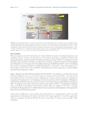
                Figure 8. For a detailed description, see the text above. Briefly, 1-at the upper right hand of the image, one sees irradiation energy
                causing breakup of the TGF-β-Ligand complex with activation of TGF-β. 2- The TGF-β receptor is activated and 3- the SMAD complex
                initiates nuclear DNA transcription that results in 4 Secretion of various factors that affect various cellular behaviors (a, b, c, d, e, f),
                culminating in a fibrotic state. 5-When Fat Grafts are injected into the fibrotic tissue, SVF MSCs within the graft release HGF, 6- HGF
                receptor is activated and 7- the GAB1 intracellular moiety initiates a group of intracellular pathways that constitute the 8- “Mitotic
                Growth Program” (a, b, c, d, e, f) that in turn causes tissue regeneration and blocks TGF-β actions.

               HGF and MSCs
               When one maps out the HGF “instruction sets”, they look like the behavior of a Mesenchymal Stem cell,
               including a feature we have not discussed yet: the immune modulatory capabilities of HGF. HGF
               immunomodulatory pathways parallel in detail the mechanism of action of MSCs. For example, both HGF
               and MSCs generate immune modulatory programs by converting “classically activated” M1 Macrophages
               (pro-inflammatory) to M2 regenerative macrophage phenotype. Both HGF and MSCs work by stimulating
               the secretion of immunomodulatory amplifying signals IL-10 and PGE-2. They both suppress pro-
               inflammatory cell lines such as Th1, Dendritic cells and promote immune suppressive Treg and MDSCs
                                             [37]
               (Myeloid Derived Suppressor Cells) .

               Figure 9 illustrates the almost identical behavior of HGF and MSCs. The diagram on the left comes from an
               article by Nakamura et al., one of the first authors to isolate and describe HGF . One can see the
                                                                                        [31]
               intracellular moiety of the HGF Receptor triggering the “mitotic growth program” including (A)
               mitogenesis; (B) angiogenesis; (C) Morphogenesis; (D) anti-apoptosis, and (E) motogenesis. Although not
               depicted in the illustration, the same article also notes the immunomodulatory and antifibrotic properties of
               HGF. The diagram on the right comes from a nature review article on MSCs . The parallels are strikingly
                                                                                [38]
               close to identical to Nakamura’s observations on HGF. MSCs are capable of (A) “Met”- (enhance
               proliferation); (B) angiogenesis; (C) differentiation; (D) anti-apoptosis; and (E) migration. The diagram also
               depicts the anti-inflammatory properties.


               At the time of publication of the articles whose illustrations are compared above, there was little
               experimental corroboration of the theory that HGF is the “master regulator” for the MSC regenerative
               response in general, and RIF specifically. But now, several lines of evidence point strongly to this
               conclusion.

