
Page 58 - Read Online
P. 58
vonderEmbse et al. Neuroimmunol Neuroinflammation 2020;7:345-59 I http://dx.doi.org/10.20517/2347-8659.2019.29 Page 353
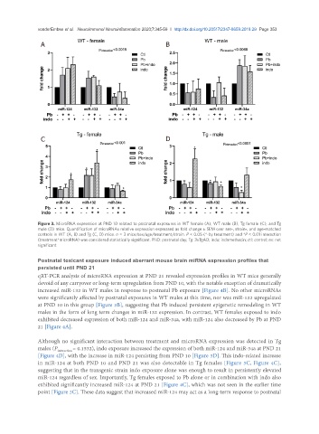
A B
C D
Figure 3. MicroRNA expression at PND 10 related to postnatal exposures in WT female (A), WT male (B), Tg female (C), and Tg
male (D) mice. Quantification of microRNAs relative expression expressed as fold change ± SEM over sex-, strain-, and age-matched
controls in WT (A, B) and Tg (C, D) mice. n = 3 mice/sex/age/treatment/strain. P < 0.05 (*-by treatment) and *P < 0.01 interaction
(treatment*microRNA) was considered statistically significant. PND: postnatal day; Tg: 3xTgAD; indo: indomethacin; ctl: control; ns: not
significant
Postnatal toxicant exposure induced aberrant mouse brain miRNA expression profiles that
persisted until PND 21
qRT-PCR analysis of microRNA expression at PND 21 revealed expression profiles in WT mice generally
devoid of any carryover or long-term upregulation from PND 10, with the notable exception of dramatically
increased miR-132 in WT males in response to postnatal Pb exposure [Figure 4B]. No other microRNAs
were significantly affected by postnatal exposures in WT males at this time, nor was miR-132 upregulated
at PND 10 in this group [Figure 3B], suggesting that Pb induced persistent epigenetic remodeling in WT
males in the form of long term changes in miR-132 expression. In contrast, WT females exposed to indo
exhibited decreased expression of both miR-124 and miR-34a, with miR-124 also decreased by Pb at PND
21 [Figure 4A].
Although no significant interaction between treatment and microRNA expression was detected in Tg
males (P interaction = 0.1532), indo exposure increased the expression of both miR-124 and miR-34a at PND 21
[Figure 4D], with the increase in miR-124 persisting from PND 10 [Figure 3D]. This indo-related increase
in miR-124 at both PND 10 and PND 21 was also detectable in Tg females [Figure 3C, Figure 4C],
suggesting that in the transgenic strain indo exposure alone was enough to result in persistently elevated
miR-124 regardless of sex. Importantly, Tg females exposed to Pb alone or in combination with indo also
exhibited significantly increased miR-124 at PND 21 [Figure 4C], which was not seen in the earlier time
point [Figure 3C]. These data suggest that increased miR-124 may act as a long-term response to postnatal