
Page 55 - Read Online
P. 55
Page 350 vonderEmbse et al. Neuroimmunol Neuroinflammation 2020;7:345-59 I http://dx.doi.org/10.20517/2347-8659.2019.29
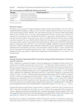
Table 1. Primer sequences of miRCURY LNA® PCR Primer sets (Exiqon)
MicroRNA RT primer sequence 5'-3'
hsa-miR-124-3p UAAGGCACGCGGUGAAUGCC target
mmu-miR-132-5p AACCGUGGCUUUCGAUUGUUAC target
hsa-miR-34a-5p UGGCAGUGUCUUAGCUGGUUGU target
SNORD110 [UGACUUAUAUAUCUGUCAAUCCCCUGAGAGAUCACUGACGACUCCAUGUGUCUGAGCAA] reference
UniSp6 CUAGUCCGAUCUAAGUCUUCGA control
RT: reverse transcriptase
Statistical analysis
All statistics were carried out using the Statistical Analysis System (SAS Institute, Cary, NC, USA) or
GraphPad PRISM (GraphPad, La Jolla, CA, USA) software. Initially, exploratory analyses were carried out
using mixed modeling (PROC MIXED, SAS) with random intercepts to evaluate possible relationships
between GxE variables with sex and age. Immunohistochemical (IHC) analyses were conducted in
untreated mice stratified by strain modeling the fixed effects of age and sex. The fixed effects of treatment
and age were then modeled, stratified by strain and sex. Possible interactions between these variables
were also evaluated. Two-way ANOVA (PROC GLM, SAS) for age and sex was performed for each
strain and treatment group for quantitative real-time polymerase chain reaction (qRT-PCR). Individual
pairwise comparisons were made with a t-test corrected for multiple comparisons using the Holm-Sidak
method or a Tukey’s studentized range distribution method. IHC data are represented as % mean DAP12
immunopositive staining/ROI over strain- and sex-matched controls at PND10 ± SEM, and qRT-PCR
data are represented as mean fold change over sex-, strain-, and age-matched controls, with respect to
endogenous reference gene levels, ± SEM. Statistical significance was determined at *P < 0.05 and *P < 0.01
for interactions.
RESULTS
Sexually dimorphic hippocampal DAP12 expression during postnatal development is altered by
early-life exposures
DAP12 expression on perinatal microglia is critical for phagocytosis of apoptotic neurons, and thus the
development of healthy neuroimmune interactions [19,49] . To determine baseline expression of DAP12
during the postnatal period in this GxE mouse model immunohistochemical analysis was quantified in
the hippocampus of untreated control mice, stratified by sex and genetic strain. In both WT and Tg males
DAP12+ expression along the dentate gyrus was not significantly altered by age [Figure 1]. In WT females,
DAP12 expression was significantly increased at both PND 15 and PND21 compared to expression levels
at PND 10 [Figure 1A and C]. Similarly, untreated Tg females had increased DAP12 by PND 21 compared
to PND 10 but was not significantly altered at PND 15 [Figure 1B and D]. Notably, while significant sex
differences were detectable in WT mice at both PND 15 and 21 [Figure 1A and C], only PND 21 differed
significantly by sex for DAP12 expression in the Tg mouse strain [Figure 1B and D]. These data suggest
that DAP12 expression in the hippocampus during the postnatal period is sexually dimorphic regardless of
genetic background and that this sexual dimorphism is dependent on age in WT mice (*P interaction < 0.0001).
Postnatal Pb exposure significantly increased DAP12 expression in both WT and Tg female mice at
PND 10 that then decreased over time [Figure 2A and C], with an inverse age-related trend compared to
controls, suggesting a profound and persistent effect on female hippocampal DAP12 expression due to Pb.
Interestingly, Tg males exposed to Pb also had significantly increased DAP12 at PND 10 that persisted with
age [Figure 2D], whereas WT male expression was unaffected by Pb at all ages [Figure 2B].
Importantly, the addition of indomethacin before Pb exposure was able to moderate the long-term
depression of DAP12 by Pb in Tg females [Figure 2C]. This is striking when contrasted with the nearly