
Page 56 - Read Online
P. 56
vonderEmbse et al. Neuroimmunol Neuroinflammation 2020;7:345-59 I http://dx.doi.org/10.20517/2347-8659.2019.29 Page 351
A B
C D
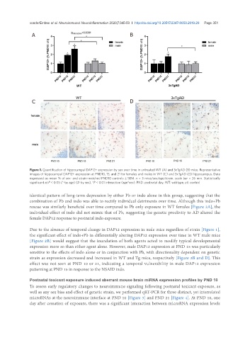
Figure 1. Quantification of hippocampal DAP12+ expression by sex over time in untreated WT (A) and 3xTgAD (B) mice. Representative
images of hippocampal DAP12+ expression at PND10, 15, and 21 for females and males in WT (C) and 3xTgAD (D) hippocampus. Data
expressed as mean % of sex- and strain-matched PND10 controls ± SEM. n = 3 mice/sex/age/strain. scale bar = 26 mm. Statistically
significant at P < 0.05 (*-by age) (#-by sex), *P < 0.01 interaction (age*sex). PND: postnatal day; WT: wildtype; ctl: control
identical pattern of long-term depression by either Pb or indo alone in this group, suggesting that the
combination of Pb and indo was able to rectify individual detriments over time. Although this indo+Pb
rescue was similarly beneficial over time compared to Pb only exposure in WT females [Figure 2A], the
individual effect of indo did not mimic that of Pb, suggesting the genetic proclivity to AD altered the
female DAP12 response to postnatal indo exposure.
Due to the absence of temporal change in DAP12 expression in male mice regardless of strain [Figure 1],
the significant effect of indo+Pb in differentially altering DAP12 expression over time in WT male mice
[Figure 2B] would suggest that the inundation of both agents acted to modify typical developmental
expression more so than either agent alone. However, male DAP12 expression at PND 15 was particularly
sensitive to the effects of indo alone or in conjunction with Pb, with directionality dependent on genetic
strain as expression decreased and increased in WT and Tg mice, respectively [Figure 2B and D]. This
effect was not seen at PND 10 or 21, indicating a temporal vulnerability in male DAP12 expression
patterning at PND 15 in response to the NSAID indo.
Postnatal toxicant exposure induced aberrant mouse brain miRNA expression profiles by PND 10
To assess early regulatory changes to neuroimmune signaling following postnatal toxicant exposure, as
well as any sex bias and effect of genetic strain, we performed qRT-PCR for three distinct, yet interrelated
microRNAs at the neuroimmune interface at PND 10 [Figure 3] and PND 21 [Figure 4]. At PND 10, one
day after cessation of exposure, there was a significant interaction between microRNA expression levels