
Page 27 - Read Online
P. 27
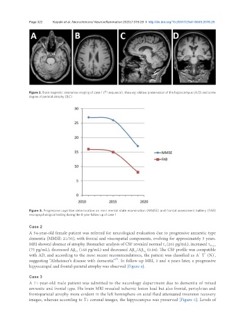
Page 322            Kapaki et al. Neuroimmunol Neuroinflammation 2020;7:319-29  I  http://dx.doi.org/10.20517/2347-8659.2019.26


               Figure 2. Brain magnetic resonance imaging of case 1 (T1 sequence), showing relative preservation of the hippocampus (A,D) and some
               degree of parietal atrophy (B,C)


               Figure 3. Progressive cognitive deterioration on mini mental state examination (MMSE) and frontal assessment battery (FAB)
               neuropsychological testing during the 8-year follow-up of case 1


               Case 2
               A 54-year-old female patient was referred for neurological evaluation due to progressive amnestic type
               dementia (MMSE: 21/30), with frontal and visuospatial components, evolving for approximately 5 years.
               MRI showed absence of atrophy. Biomarker analysis of CSF revealed normal τ (261 pg/mL), increased τ P-181
                                                                                 T
               (75 pg/mL), decreased Aβ  (168 pg/mL) and decreased Aβ /Aβ  (0.04). The CSF profile was compatible
                                                                       40
                                      42
                                                                  42
                                                                                                  +
               with AD, and according to the most recent recommendations, the patient was classified as A  T  (N) ,
                                                                                                         -
                                                                                                    +
                                                         [7]
               suggesting “Alzheimer’s disease with dementia” . In follow-up MRI, 3 and 4 years later, a progressive
               hippocampal and frontal-parietal atrophy was observed [Figure 4].
               Case 3
               A 71-year-old male patient was admitted to the neurology department due to dementia of mixed
               amnestic and frontal type. His brain MRI revealed ischemic lesion load but also frontal, perisylvian and
               frontoparietal atrophy more evident in the left hemisphere on axial fluid attenuated inversion recovery
               images, whereas according to T1 coronal images, the hippocampus was preserved [Figure 5]. Levels of