
Page 28 - Read Online
P. 28
Kapaki et al. Neuroimmunol Neuroinflammation 2020;7:319-29 I http://dx.doi.org/10.20517/2347-8659.2019.26 Page 323
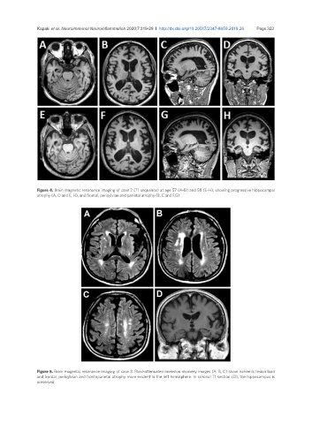
Figure 4. Brain magnetic resonance imaging of case 2 (T1 sequence) at age 57 (A-D) and 58 (E-H), showing progressive hippocampal
atrophy (A, D and E, H), and frontal, perisylvian and parietal atrophy (B, C and F,G)
Figure 5. Brain magnetic resonance imaging of case 3. Fluid-attenuated inversion recovery images (A, B, C) show ischemic lesion load
and frontal, perisylvian and frontoparietal atrophy more evident in the left hemisphere. In coronal T1 section (D), the hippocampus is
preserved