
Page 29 - Read Online
P. 29
Page 324 Kapaki et al. Neuroimmunol Neuroinflammation 2020;7:319-29 I http://dx.doi.org/10.20517/2347-8659.2019.26
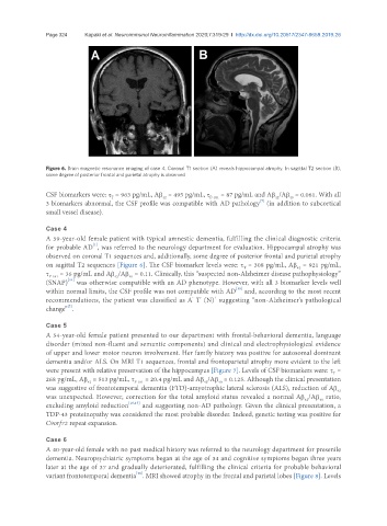
Figure 6. Brain magnetic resonance imaging of case 4. Coronal T1 section (A) reveals hippocampal atrophy. In sagittal T2 section (B),
some degree of posterior frontal and parietal atrophy is observed
CSF biomarkers were: τ = 963 pg/mL, Aβ = 495 pg/mL, τ P-181 = 87 pg/mL and Aβ /Aβ = 0.061. With all
42
42
40
T
[7]
3 biomarkers abnormal, the CSF profile was compatible with AD pathology (in addition to subcortical
small vessel disease).
Case 4
A 59-year-old female patient with typical amnestic dementia, fulfilling the clinical diagnostic criteria
[1]
for probable AD , was referred to the neurology department for evaluation. Hippocampal atrophy was
observed on coronal T1 sequences and, additionally, some degree of posterior frontal and parietal atrophy
on sagittal T2 sequences [Figure 6]. The CSF biomarker levels were: τ = 308 pg/mL, Aβ = 921 pg/mL,
42
T
τ P-181 = 36 pg/mL and Aβ /Aβ = 0.11. Clinically, this “suspected non-Alzheimer disease pathophysiology”
40
42
(SNAP) was otherwise compatible with an AD phenotype. However, with all 3 biomarker levels well
[14]
[15]
within normal limits, the CSF profile was not compatible with AD and, according to the most recent
+
-
-
recommendations, the patient was classified as A T (N) suggesting “non-Alzheimer’s pathological
[7]
change” .
Case 5
A 54-year-old female patient presented to our department with frontal-behavioral dementia, language
disorder (mixed non-fluent and semantic components) and clinical and electrophysiological evidence
of upper and lower motor neuron involvement. Her family history was positive for autosomal dominant
dementia and/or ALS. On MRI T1 sequences, frontal and frontoparietal atrophy more evident to the left
were present with relative preservation of the hippocampus [Figure 7]. Levels of CSF biomarkers were: τ =
T
268 pg/mL, Aβ = 513 pg/mL, τ P-181 = 20.4 pg/mL and Aβ /Aβ = 0.125. Although the clinical presentation
42
42
40
was suggestive of frontotemporal dementia (FTD)-amyotrophic lateral sclerosis (ALS), reduction of Aβ
42
was unexpected. However, correction for the total amyloid status revealed a normal Aβ /Aβ ratio,
42
40
excluding amyloid reduction [16,17] and suggesting non-AD pathology. Given the clinical presentation, a
TDP-43 proteinopathy was considered the most probable disorder. Indeed, genetic testing was positive for
C9orf72 repeat expansion.
Case 6
A 40-year-old female with no past medical history was referred to the neurology department for presenile
dementia. Neuropsychiatric symptoms began at the age of 34 and cognitive symptoms began three years
later at the age of 37 and gradually deteriorated, fulfilling the clinical criteria for probable behavioral
[18]
variant frontotemporal dementia . MRI showed atrophy in the frontal and parietal lobes [Figure 8]. Levels