
Page 244 - Read Online
P. 244
Page 6 of 10 Raevsky et al. Neuroimmunol Neuroinflammation 2018;5:33 I http://dx.doi.org/10.20517/2347-8659.2018.34
A B C
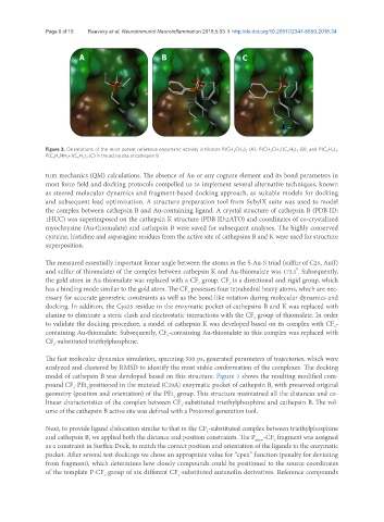
Figure 3. Orientations of the most potent reference enzymatic activity inhibitors P(CH 2 CH 3 ) 3 (A), P(CH 2 CH 3 )(C 6 H 5 ) 2 (B), and P(C 6 H 5 ) 3 ,
P(C 6 H 4 NH 3 +)(C 6 H 5 ) 2 (C) in the active site of cathepsin B
tum mechanics (QM) calculations. The absence of Au or any cognate element and its bond parameters in
most force field and docking protocols compelled us to implement several alternative techniques, known
as steered molecular dynamics and fragment-based docking approach, as suitable models for docking
and subsequent lead optimization. A structure preparation tool from SybylX suite was used to model
the complex between cathepsin B and Au-containing ligand. A crystal structure of cathepsin B (PDB ID:
1HUC) was superimposed on the cathepsin K structure (PDB ID:2ATO) and coordinates of co-crystalized
myochrysine (Au-thiomalate) and cathepsin B were saved for subsequent analyses. The highly conserved
cysteine, histidine and asparagine residues from the active site of cathepsins B and K were used for structure
superposition.
The measured essentially important linear angle between the atoms in the S-Au-S triad (sulfur of C25, Au(I)
o
and sulfur of thiomalate) of the complex between cathepsin K and Au-thiomalate was 173.1 . Subsequently,
the gold atom in Au-thiomalate was replaced with a CF group. CF is a directional and rigid group, which
3
3
has a binding mode similar to the gold atom. The CF possesses four tetrahedral heavy atoms, which are nec-
3
essary for accurate geometric constraints as well as the bond-like rotation during molecular dynamics and
docking. In addition, the Cys25 residue in the enzymatic pocket of cathepsins B and K was replaced with
alanine to eliminate a steric clash and electrostatic interactions with the CF group of thiomalate. In order
3
to validate the docking procedure, a model of cathepsin K was developed based on its complex with CF -
3
containing Au-thiomalate. Subsequently, CF -containing Au-thiomalate in this complex was replaced with
3
CF -substituted triethylphosphine.
3
The fast molecular dynamics simulation, spanning 500 ps, generated parameters of trajectories, which were
analyzed and clustered by RMSD to identify the most stable conformation of the complexes. The docking
model of cathepsin B was developed based on this structure. Figure 3 shows the resulting modified com-
pound CF -PEt positioned in the mutated (C29A) enzymatic pocket of cathepsin B, with preserved original
3
3
geometry (position and orientation) of the PEt group. This structure maintained all the distances and co-
3
linear characteristics of the complex between CF -substituted triethylphosphine and cathepsin B. The vol-
3
ume of the cathepsin B active site was defined with a Protomol generation tool.
Next, to provide ligand dislocation similar to that in the CF -substituted complex between triethylphosphine
3
and cathepsin B, we applied both the distance and position constraints. The P -CF fragment was assigned
3
atom
as a constraint in Surflex-Dock, to match the correct position and orientation of the ligands in the enzymatic
pocket. After several test dockings we chose an appropriate value for “cpen” function (penalty for deviating
from fragment), which determines how closely compounds could be positioned to the source coordinates
of the template P-CF group of six different CF -substituted auranofin derivatives. Reference compounds
3
3