
Page 243 - Read Online
P. 243
Raevsky et al. Neuroimmunol Neuroinflammation 2018;5:33 I http://dx.doi.org/10.20517/2347-8659.2018.34 Page 5 of 10
A B
C D
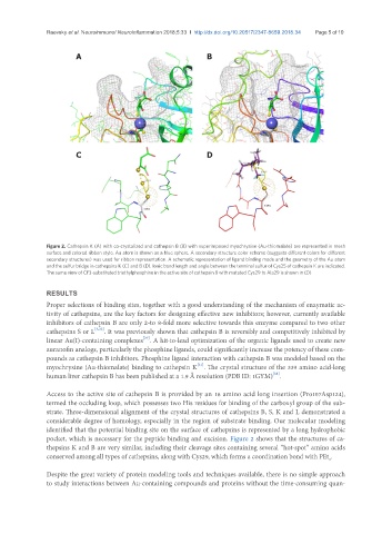
Figure 2. Cathepsin K (A) with co-crystalized and cathepsin B (B) with superimposed myochrysine (Au-thiomalate) are represented in mesh
surface and colored ribbon style. Au atom is shown as a blue sphere. A secondary structure color scheme (suggests different colors for different
secondary structures) was used for ribbon representation. A schematic representation of ligand binding mode and the geometry of the Au atom
and the sulfur bridge in cathepsins K (C) and B (D). Ionic bond length and angle between the terminal sulfur of Cys25 of cathepsin K are indicated.
The same view of CF3-substituted triethylphosphine in the active site of cathepsin B with mutated Cys29 to Ala29 is shown in (D)
RESULTS
Proper selections of binding sites, together with a good understanding of the mechanism of enzymatic ac-
tivity of cathepsins, are the key factors for designing effective new inhibitors; however, currently available
inhibitors of cathepsin B are only 2-to 8-fold more selective towards this enzyme compared to two other
cathepsins S or L [3,22] . It was previously shown that cathepsin B is reversibly and competitively inhibited by
linear Au(I)-containing complexes . A hit-to-lead optimization of the organic ligands used to create new
[29]
auranofin analogs, particularly the phosphine ligands, could significantly increase the potency of these com-
pounds as cathepsin B inhibitors. Phosphine ligand interaction with cathepsin B was modeled based on the
[31]
myochrysine (Au-thiomalate) binding to cathepsin K . The crystal structure of the 339 amino acid-long
[38]
human liver cathepsin B has been published at a 1.9 Å resolution (PDB ID: 1GYM) .
Access to the active site of cathepsin B is provided by an 18 amino acid long insertion (Pro107 Asp124),
termed the occluding loop, which possesses two His residues for binding of the carboxyl group of the sub-
strate. Three-dimensional alignment of the crystal structures of cathepsins B, S, K and L demonstrated a
considerable degree of homology, especially in the region of substrate binding. Our molecular modeling
identified that the potential binding site on the surface of cathepsins is represented by a long hydrophobic
pocket, which is necessary for the peptide binding and excision. Figure 2 shows that the structures of ca-
thepsins K and B are very similar, including their cleavage sites containing several “hot-spot” amino acids
conserved among all types of cathepsins, along with Cys29, which forms a coordination bond with PEt .
3
Despite the great variety of protein modeling tools and techniques available, there is no simple approach
to study interactions between Au-containing compounds and proteins without the time-consuming quan-