
Page 83 - Read Online
P. 83
Etemadifar et al. Imaging of demyelinated corpus callosum
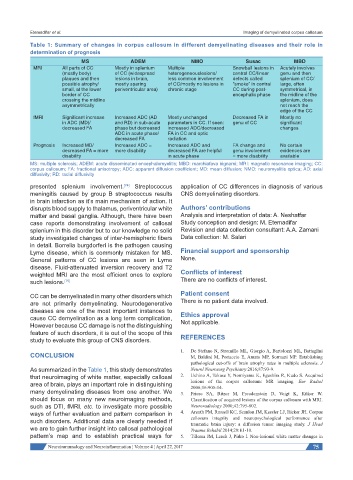
Table 1: Summary of changes in corpus callosum in different demyelinating diseases and their role in
determination of prognosis
MS ADEM NMO Susac MBD
MRI All parts of CC Mostly in splenium Multiple Snowball lesions in Acutely involves
(mostly body) of CC (widespread heterogeneouslesions/ central CC/linear genu and then
plaques and then lesions in brain, less common involvement defects called splenium of CC/
possible atrophy/ mostly sparing of CC/mostly no lesions in “smoke” in central large, often
small, at the lower periventricular area) chronic stage CC during post- symmetrical, in
border of CC encephalic phase the midline of the
crossing the midline splenium, does
asymmetrically not reach the
edge of the CC
fMRI Significant increase Increased ADC (AD Mostly unchanged Decreased FA in Mostly no
in ADC (MD)/ and RD) in sub-acute parameters in CC. If seen: genu of CC significant
decreased FA phase but decreased increased ADC/decreased changes
ADC in acute phase/ FA in CC and optic
decreased FA radiation
Prognosis Increased MD/ Increased ADC = Increased ADC and FA change and No certain
decreased FA = more more disability decreased FA are helpful genu involvement evidences are
disability in acute phase = more disability available
MS: multiple sclerosis; ADEM: acute disseminated encephalomyelitis; MBD: marchiafava bignami; MRI: magnetic resonance imaging; CC:
corpus callosum; FA: fractional anisotropy; ADC: apparent diffusion coefficient; MD: mean diffusion; NMO: neuromyelitis optica; AD: axial
diffusivity; RD: radial diffusivity
presented splenium involvement. Streptococcus application of CC differences in diagnosis of various
[74]
meningitis caused by group B streptococcus results CNS demyelinating disorders.
in brain infarction as it’s main mechanism of action. It
disrupts blood supply to thalamus, periventricular white Authors’ contributions
matter and basal ganglia. Although, there have been Analysis and interpretation of data: A. Neshatfar
case reports demonstrating involvement of callosal Study conception and design: M. Etemadifar
splenium in this disorder but to our knowledge no solid Revision and data collection consultant: A.A. Zamani
study investigated changes of inter-hemispheric fibers Data collection: M. Salari
in detail. Borrelia burgdorferi is the pathogen causing
Lyme disease, which is commonly mistaken for MS. Financial support and sponsorship
General patterns of CC lesions are seen in Lyme None.
disease. Fluid-attenuated inversion recovery and T2
weighted MRI are the most efficient ones to explore Conflicts of interest
such lesions. [75] There are no conflicts of interest.
CC can be demyelinated in many other disorders which Patient consent
are not primarily demyelinating. Neurodegenerative There is no patient data involved.
diseases are one of the most important instances to Ethics approval
cause CC demyelination as a long term complication,
However because CC damage is not the distinguishing Not applicable.
feature of such disorders, it is out of the scope of this
study to evaluate this group of CNS disorders. REFERENCES
1. De Stefano N, Stromillo ML, Giorgio A, Bartolozzi ML, Battaglini
CONCLUSION M, Baldini M, Portaccio E, Amato MP, Sormani MP. Establishing
pathological cut-offs of brain atrophy rates in multiple sclerosis. J
As summarized in the Table 1, this study demonstrates Neurol Neurosurg Psychiatry 2016;87:93-9.
that neuroimaging of white matter, especially callosal 2. Uchino A, Takase Y, Nomiyama K, Egashira R, Kudo S. Acquired
area of brain, plays an important role in distinguishing lesions of the corpus callosum: MR imaging. Eur Radiol
2006;16:905-14.
many demyelinating diseases from one another. We 3. Friese SA, Bitzer M, Freudenstein D, Voigt K, Küker W.
should focus on many new neuroimaging methods, Classification of acquired lesions of the corpus callosum with MRI.
such as DTI, fMRI, etc. to investigate more possible Neuroradiology 2000;42:795-802.
ways of further evaluation and pattern comparison in 4. Arenth PM, Russell KC, Scanlon JM, Kessler LJ, Ricker JH. Corpus
such disorders. Additional data are clearly needed if callosum integrity and neuropsychological performance after
traumatic brain injury: a diffusion tensor imaging study. J Head
we are to gain further insight into callosal pathological Trauma Rehabil 2014;29:E1-10.
pattern’s map and to establish practical ways for 5. Tillema JM, Leach J, Pirko I. Non-lesional white matter changes in
Neuroimmunology and Neuroinflammation ¦ Volume 4 ¦ April 27, 2017 75