
Page 87 - Read Online
P. 87
Chen et al. Anti-N-methyl-d-aspartate receptor autoimmune encephalitis with sensory attack
INTRODUCTION Repeated lumbar puncture showed no significant
change in cell count, glucose, or protein level.
Limbic encephalitis is believed to be a disorder Herpes simplex virus (HSV) and cytomegalovirus
affecting the medial temporal lobe of the brain. The (CMV) polymerase chain reactions were negative
underlying cause can be either of autoimmune origin in cerebrospinal fluid (CSF). The immunoglobulin G
or viral infection. N-methyl-d-aspartate-receptor (IgG) index was 0.5. CSF oligoclonal banding was also
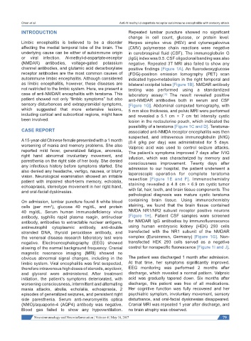
(NMDAR) antibodies, voltage-gated potassium negative. Repeated 3T MRI also failed to show any
channel antibodies, and glutamic acid decarboxylase positive findings [Figure 1A]. An fluorodeoxyglucose
receptor antibodies are the most common causes of (FDG)-positron emission tomography (PET) scan
autoimmune limbic encephalitis. Although considered indicated hypo-metabolism in the right temporal and
as limbic encephalitis, however, these diseases are bilateral occipital lobes [Figure 1B]. NMDAR antibody
not restricted to the limbic system. Here, we present a testing was performed using a standardized
case of anti-NMDAR encephalitis with teratoma. This laboratory assay. The result revealed positive
[1]
patient showed not only “limbic symptoms” but also anti-NMDAR antibodies both in serum and CSF
sensory disturbances and extrapyramidal symptoms, [Figure 1G]. Abdominal computed tomography, with
which suggested that more extensive lesions, 5 mm slice thickness, and pelvic MRI were performed
including cortical and subcortical regions, might have and revealed a 5.1 cm × 7 cm fat intensity cystic
been involved. lesion in the rectouterine pouch, which indicated the
possibility of a teratoma [Figure 1C and D]. Teratoma-
CASE REPORT associated anti-NMDA receptor encephalitis was then
suspected, and intravenous immunoglobulin (IVIG)
A 15-year-old Chinese female presented with a 1 month (0.4 g/kg per day) was administrated for 5 days.
worsening of mania and memory problems. She also Valproic acid was used to control seizure attacks.
reported mild fever, generalized fatigue, anorexia, The patient’s symptoms improved 7 days after IVIG
right hand abnormal involuntary movement, and infusion, which was characterized by memory and
paresthenia on the right side of her body. She denied consciousness improvement. Twenty days after
any infectious history before symptoms started. She admission to our hospital, the patient underwent a
also denied any headache, vertigo, nausea, or blurry laparoscopic operation for complete teratoma
vision. Neurological examination showed an irritable resection [Figure 1E and F]. Immunochemistry
patient with impaired short-term memory, echolalia,
echoapraxia, stereotype movement in her right hand, staining revealed a 4.8 cm × 6.9 cm cystic tumor
and oral-facial dyskinesias. with fat, hair, teeth, and brain tissue components. The
pathological diagnosis was mature cystic teratoma
On admission, lumbar puncture found 8 white blood containing brain tissue. Using immunochemistry
cells (per mm ), glucose 40 mg/dL, and protein staining, we found that the brain tissue contained
3
40 mg/dL. Serum human immunodeficiency virus NMDA NR1/NR2 subunit receptor positive neurons
antibody, syphilis rapid plasma reagin, antinuclear [Figure 1H]. Patient CSF samples were screened
antibody, antibodies to extractable nuclear antigens, for NMDAR IgG antibodies by immunofluorescence
antineutrophil cytoplasmic antibody, anti-double using human embryonic kidney (HEK) 293 cells
stranded DNA, thyroid peroxidase antibody, and transfected with the NR1 subunit of the NMDAR
the venereal disease research laboratory test were complex (Euroimmun, Germany) [Figure 1G]. Non-
negative. Electroencephalography (EEG) showed transfected HEK 293 cells served as a negative
slowing of the normal background frequency. Cranial control for nonspecific fluorescence [Figure 1I and J].
magnetic resonance imaging (MRI) showed no
obvious abnormal signal changes, including in the The patient was discharged 1 month after admission.
limbic system. Viral encephalitis was first suspected, At that time, her symptoms significantly improved.
therefore intravenous high doses of steroids, acyclovir, EEG monitoring was performed 2 months after
and glycerol were administered. After treatment discharge, which revealed a normal pattern. Valproic
initiation, the patient’s symptoms deteriorated, with acid was gradually tapered down. Six months after
worsening consciousness, intermittent and alternating discharge, this patient was free of all medications.
mania attacks, abulia, echolalia, echoapraxia, 2 Her cognitive function was fully recovered and her
episodes of generalized seizures, and persistent right psychiatric symptom, involuntary movement, sensory
side paresthenia. Serum anti-neuromyelitis optica disturbance, and oral-facial dyskinesias disappeared.
(NMO)/aquaporin-4 (AQP4) antibody was negative. Cranial MRI was repeated 1 year after discharge, and
Blood gas failed to show any hypoventilation. no brain atrophy was observed.
Neuroimmunology and Neuroinflammation ¦ Volume 4 ¦ May 10, 2017 79