
Page 240 - Read Online
P. 240
Li et al. Sparganosis of the brain
were various types of seizures. In some cases, it can
A B
even present as an intracranial hemorrhage . Fever
[19]
is not a common symptom. Lesions are mainly located
at frontal-partial lobes but invasion to cerebellums
are also reported in very few cases [18] . Diagnosis of
cerebral sparganosis is relatively hard since it has
no specific manifestations. A history of traveling or
living in endemic areas may indicate a possibility for
the diagnosis. In China, the majority infected people
had a history of consuming undercooked frog or
snake meat. Besides histories of traveling or living
in endemic areas, an active infection of other organs
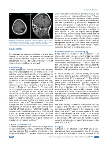
Figure 2: Postoperative enhanced T1WI MRI showed that the lesion is also a useful clue for the diagnosis of cerebral
was totally resected with a slight edmea around the operative area, A: sparganosis. In our case, the patient had a migratory
axial view; B: sagittal view. MRI: magnetic resonance imaging
nodule on the right upper limb, which makes us highly
suspect a diagnosis of cerebral sparganosis.
DISCUSSION
Laboratory test and neuroimaging
To investigate the epidemic and clinical characteristics Blood can show an increase of eosinophilia [13] . The
of cerebral sparganosis. We searched Pubmed using disease can also be diagnosed with antigen specified
the keywords “cerebral sparganosis” and “cerebral IgG antibodies from blood and CSF samples,
sparganosis mansonoide”. English literatures with at although cross-reactivity with other infestations or
least abstract available were reviewed. clonorchiasis limititsspecificity [13,18] . In our case, the
first CSF sample was negative for immune-reactivity
Epidemiology but positive for blood sample, however, the second
Human sparganosis usually occurs when patients blood and CSF sample were both positive.
consume undercooked frog or snake meat, drink
unboiled water contaminated by procercoidlarve. In CT scans usually show a mixed density lesion with
some rural areas, people use fresh snake or frog peri-lesion edema, punctate calcification is shown
meat directly on open wounds as a cure method. in approximately 50% of patients [15,20] . The MRI of
Sparganosis can be transmitted through this way, cerebral sparganosis is difficult to identify from low
[5]
too . Cases of human sparganosis are mostly grade glioma or other tumors. Typically, ring-like or
[5]
reported in east Asia, especially in China , South string-knot enhancement in T1WI images shows
[3]
Korea [15] , Thailand and Japan [16] . Till now, only the sign of sparganosis movement in the brain
three cerebral sparganosis cases were reported parenchyma. Other characteristic features include
in Europe [9,17] . Sparganosis can migrate to various a tunnel-shape configuration due to immigration of
tissues and organs, such as subcutaneous connective sparganosis through the brain parenchyma. Moreover,
tissue, muscle, breast, lungs and the central nervous serial imaging may demonstrate the tunnel sign from a
system . Reported cerebral sparganosis cases are small nodular lesion and reflect the sparganosis activity
[2]
quite small compared to other site sparganosis, but in the brain [21-23] .
[18]
according to Hong et al. the prevalence of cerebral
sparganosis was underestimated, many cases were MR spectroscopy of cerebral sparganosis (Bo and
not reported. In addition, a lot of Asian patients were Xuejian [24] and Chiu et al. [25] ) showed increased Cho
reported with local language rather than English. The and decreased NAA peaks in their cases, as in our
incidence of cerebral sparganosis of children and case. Chiu et al. [25] also revealed peaks at 1.3 ppm
young people are higher than adults, and people in and a peak between 14 and 1.8 ppm in his 46-year-
rural counties who are in underprivileged financial old female patient, however, our case did not show
status have a great opportunity to get a sparganosis a similar result. MR perfusion did not show higher
infection due to hygiene problems . cerebral perfusion in our case that was seen in the
[13]
case of Chiu et al. [25] .
Clinical characteristics
Cerebral sparganosis can cause various symptoms Surgery
including headache, seizure, parenthesis, confusion, Although some studies report that using anti-
memory loss, etc. The symptoms are related to the sparganosis drugs can successfully cure the disease,
[14]
location of the lesion , the most common symptoms the most efficient way to cure the disease still is
240 Neuroimmunology and Neuroinflammation ¦ Volume 4 ¦ November 20, 2017