
Page 239 - Read Online
P. 239
Li et al. Sparganosis of the brain
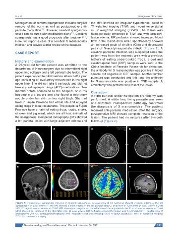
Management of cerebral sparganosis includes surgical the MRI showed an irregular hypointense lesion in
removal of the worm as well as postoperative anti- T1 weighted imaging (T1WI) and hyperintense signal
[13]
parasite medication . However, cerebral sparganosis in T2 weighted imaging (T2WI). The lesion was
[14]
cases can be cured with medication alone . Cerebral homogenously enhanced in T1WI and with largeperi-
[13]
sparganosis has a good prognosis after treatment . lesion edema. MR perfusion showed increased blood
Here, we report a case of a cerebral S mansonoides flow in the lesion area while spectroscopy showed
infection and provide a brief review of the literature. an increased peak of choline (Cho) and decreased
peak of N-acetyl-aspartate (NAA) [Figure 1]. A
CASE REPORT cerebral parasitic infection was suspected since the
patient was from the endemic area with a previous
history of eating undercooked frogs. Blood and
History and examination cerebrospinal fluid (CSF) samples were sent to the
A 29-year-old female patient was admitted to the China Institute of Parasite Research for detection,
department of Neurosurgery due to intermittent right the antibody for S mansonoides was positive in blood
upper limb epilepsy and a left parietal lobe lesion. The sample but negative in CSF sample. Another lumbar
patient experienced her first seizure attack half a year puncture was conducted and this time the antibody
ago consisting of involuntary movements in the right for S mansonoide was positive in CSF sample. A
upper limb. She did not take it seriously and did not craniotomy was performed to resect the lesion.
take any anti-epileptic drugs (AED) medications. Two
months before admission to the hospital, seizures Operation
became more severe and she found a migratory A right parietal under-navigation craniotomy was
nodule under her skin on her right thigh. She had performed. A white long living parasite was seen
lived in Fujian Province her whole life and enjoyed and extracted. Postoperative pathology confirmed
eating frogs in local restaurants. The people in Fujian the diagnosis of S mansonoides. The patient
Province have a habit of eating frogs, crabs, snake, received anti-parasite medication after the surgery,
chicken and pig meat, which are all host animals of postoperative MRI showed complete resection of the
the sparganosis. Computed tomography (CT) showed lesion. The patient had no seizures after 6-month
a left parietal lesion with large adjacent edema and follow-up [Figure 2].
A B C D
E F G H
Figure 1: Preoperative radiological features of cerebral sparganosis. A: axial view of CT scanning showed irregular edema of the left
parietal lobe; B: axial view of T1WI MRI showed a slight edema of the left parietal lobe; C: axial view of T2WI MRI; D: axial view of FLAIR
MRI; E: sagittal view of enhanced T1WI MRI showing an irregular enhanced lesion of the let parietal lobe; F: axial view of enhanced T1WI
MRS showed an increase of the Cho/NAA ratio; G: axial view of MR perfuison showed the lesion was hypometabolic; H: sagittal view of
preoperative DTI. CT: computed tomography; MRI: magnetic resonance imaging; NAA: N-acetyl-aspartate; T1WI: T1 weighted imaging;
DTI: diffusion tensor imaging
Neuroimmunology and Neuroinflammation ¦ Volume 4 ¦ November 20, 2017 239