
Page 233 - Read Online
P. 233
Chaurasia et al. Quadrigeminal plate lipoma
and diplopia for 2 months. He also complained of of all intracranial tumours. These benign lesions are
generalized weakness since childhood. There was no thought to arise from differentiation of the meninx
history of seizure, loss of consciousness or behavioural primitiva, a mesenchymal derivative of neural crest,
changes. On examination, it revealed normal general to lipoma tissue. The vast majority of these types of
[1]
condition having all systemic examinations within normal lesions occur near the midline . More than 50% have
except nervous system examination where we found been reported to be associated with congenital brain
diplopia, the 6th cranial nerve palsy on right side [Figure 1] malformations such as agenesis or hypoplasia of the
[2]
and bilateral papilloedema on fundoscopic examination. corpus callosum . Others include the absence of the
septum pellucidum, cranium bifidum, spina bifida,
All laboratory finding including that for fitness for being myelomeningocele, hypoplasia of the vermis and
[1]
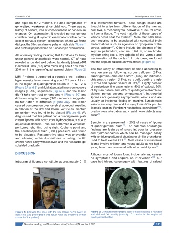
under general anaesthesia were normal. CT of head malformation of the cortex . In this case, we found
revealed a rounded well defined fat density [density-101 that the septum pallucidum was absent [Figure 4].
Hounsfield units (HU)] area measuring about 17.6 mm ×
20.6 mm in the region of quadrigeminal cistern [Figure 2]. The frequency of intracranial lipomas according to
location are as following: corpus callosum (64%),
MRI findings suggested a rounded well defined quadrigeminal-ambient cistern (13%), infundibular-
hyperintensity lesion measuring about 2.1 cm × 1.9 cm chiasmatic region (13%), cerebellopontine angle
[3]
in the region of quadrigeminal cistern in T1-W, T2-W (0.06%) and Sylvian fissure (0.03%) . Eighty percent
[Figure 3A and B] and fluid attenuated inversion recovery of cerebellopontine angle lesions, 50% of callosal, 50%
images (FLAIR) sequences [Figure 4] and the lesion of Sylvian fissure and 20% of quadrigeminal-ambient
[2]
didn’t take contrast enhancement [Figure 3C] and cistern lipomas become symptomatic . Intracranial
diffusion weighted image (DWI) sequence suggested lipomas are generally asymptomatic lesions and are
no restriction of diffusion [Figure 3D]. The lesion usually an incidental finding on imaging. Symptomatic
caused compression over cerebral aqueduct resulting lesions are very rare and the symptoms differ per the
[4,5]
in dilation of the 3rd and lateral ventricles. Septum lipoma’s location. Persistent headaches, convulsions ,
pellucidum was found to be absent [Figure 4]. We psychomotor retardation and cranial nerve defects may
[6]
diagnosised that this patient had a quadrigeminal plate occur .
cistern lipoma with obstructive hydrocephalous due to
aqueductal stenosis. Thus, we performed a ventriculo- Symptoms are presented in 20% of cases of lipoma
[7]
peritoneal shunting using right Kocher’s point and of quadrigeminal plate . The common neurologic
the cerebrospinal fluid (CSF) pressure was found findings are features of raised intracranial pressure
to be elevated. Postoperative state was uneventful and hydrocephalous which can be managed easily
and following ventriculo-peritoneal shunting, the 6th with ventriculoperitoneal shunting or similar procedures
[8]
cranial nerve palsy was resolved and the headache got used to treat excess CSF . Most cases of intracranial
subsided gradually. lipoma involve children and young adults as we had a
[9]
young teen male presented with intracranial lipoma .
DISCUSSION Although most of lipoma found incidentally and causes
[8]
no symptoms and require no intervention , our
Intracranial lipomas constitute approximately 0.1% case had triventriculomegaly with features of raised
Figure 1: Showing the case with the 6th cranial nerve palsy on Figure 2: Computed tomography scan of head showing a rounded
right side (the photograph was taken with the informed written well defined fat density (density 101) lesion in the region of
consent of the patient) quadrigeminal cistern
Neuroimmunology and Neuroinflammation ¦ Volume 4 ¦ November 9, 2017 233