
Page 152 - Read Online
P. 152
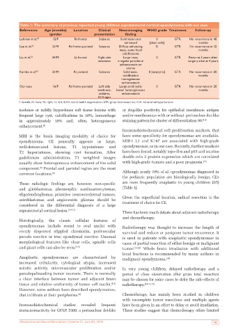
Table 1: The summary of previous reported young children supratentorial cortical ependymoma with our case.
           References   Age (months)   Location    Clinical   Neuroimaging  WHO grade Treatment    Follow-up
                          /gender                presentation    finding
           Lehman et al, [4]   12/F    Rt frontal   Seizures  Solid lesion not   III     GTR     No recurrence in 48
                                                                enhanced    [clear cells]            months
           Lee et al, [2]  21/M     Rt fronto-parietal   Seizures  Diffuse enhancing   II  GTR   No recurrence in 12
                                                             mass, some focal                        months
                                                               calcifications
           Liu et al, [1]  24/M        Lt frontal  Right side   Large mass,     III      GTR    Recur at 3 years after
                                                   weakness  irregular peripheral               surgery alive at 4 years
                                                             enhancement no
                                                                 edema
           Kambe et al, [6]  24/M     Rt parietal  Seizures    Solid mass,   II [tanycytic]  GTR  No recurrence in 20
                                                               calcification                         months
                                                              homogeneous
                                                              enhancement
           Our case         16/F    Rt fronto-parietal   Left side   Large solid cystic   III  GTR  No recurrence in 20
                                                  weakness,   lesion heterogeneous                   months
                                                   seizures,  enhancement
                                                   ICH signs
           F: female; M: male; Rt: right; Lt: left; WHO: world health organization; GTR: gross total resection; ICH: intracranial hypertension
           isodense or mildly hyperdense soft tissue lesions with   or ring-like positivity for epithelial membrane antigen
           frequent large cyst, calcifications in 50%, hemorrhage   and/or membranous with or without  perinuclear dot-like
           in approximately 10% and, often, heterogeneous      staining pattern for cluster of differentiation 99. [1,4]
           enhancement. [8]
                                                               Immunohistochemical cell proliferation markers, that
           MRI is the brain imaging modality of choice for     have some specificity for ependymomas are available.
           ependymoma. CE generally appears as large,          MIB-1 L1 and Ki-67 are associated with high-grade
           well-demarcated  lesions,  T1  hypointense   and    ependymomas, as in our case. Recently, further markers
           T2 hyperintense, showing cyst formation. After      have been found, notably topo-II-α and p53 and murine
           gadolinium administration, T1 weighted images       double min 2 protein expression which are correlated
           usually show heterogeneous enhancement of the solid   with high-grade tumors and a poor prognosis. [12]
           component.  Frontal and parietal region are the most
                     [8]
           common locations. [8]                               Although nearly 70% of all ependymomas diagnosed in
                                                               the pediatric population are histologically benign, CEs
           These radiologic findings are, however, non-specific   are more frequently anaplastic in young children (3/5)
           and  glioblastomas,  pleomorphic  xanthoastrocytomas,   [Table 1].
           oligodendrogliomas, primitive neuroectodermal tumors,
           astroblastomas and angiocentric gliomas should be   Given the superficial location, radical resection is the
           considered in the differential diagnosis of a large   treatment of choice for CE.
           supratentorial cortical lesion. [1,8-11]            There has been much debate about adjuvant radiotherapy
                                                               and chemotherapy.
           Histologically, the classic cellular features of
           ependymomas include round to oval nuclei with       Radiotherapy was thought to increase the length of
           evenly  dispersed  stippled  chromatin,  perivascular   survival and reduce or postpone tumor recurrence. It
           pseudo rosettes or true ependymal rosettes. Unusual   is used in patients with anaplastic ependymomas in
           morphological features like clear cells, spindle cells   cases of partial resection of either benign or malignant
           and giant cells can also be seen. [1,4]             tumor. [13,14]  Whole brain irradiation with additional
                                                               local fractions is recommended by many authors in
           Anaplastic ependymomas are characterized by         malignant ependymoma. [14]
           increased cellularity, cytological atypia, increased
           mitotic activity, microvascular proliferation and/or   In very young children, delayed radiotherapy and a
           pseudopalissading tumor necrosis. There is normally   period of close observation after gross total resection
           a  clear  interface  between  tumor  and  adjacent  brain   may be chosen for some cases to defer the side-effects of
           tissue and relative uniformity of tumor cell nuclei. [1,4]    radiotherapy. [4,6,13,15]
           However, some authors have described ependymomas,
           that infiltrate at their peripheries. [4]           Chemotherapy has mainly been studied in children
                                                               with incomplete tumor resections and multiple agents
           Immunohistochemical   studies  revealed  frequent   have been given in an effort to delay or avoid irradiation.
           immunoreactivity for GFAP, S100, a perinuclear dot-like   These studies suggest that chemotherapy offers limited


           Neuroimmunol Neuroinflammation | Volume 3 | June 20, 2016                                         143