
Page 151 - Read Online
P. 151
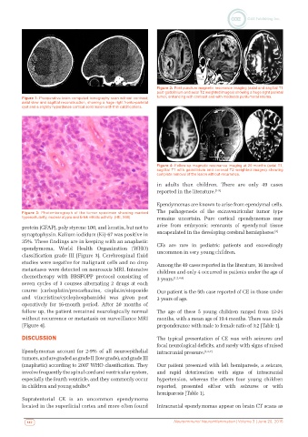
Figure 2: Post puncture magnetic resonance imaging (axial and sagittal T1
                                                              post gadolinium and axial T2 weighted images) showing a huge right parietal
          Figure 1: Preoperative brain computed tomography scan without contrast;   tumor, enhancing with contrast and with moderate peritumoral edema.
          axial view and sagittal reconstruction, showing a huge right fronto-parietal
          cyst and a slightly hyperdense cortical solid lesion with thin calcifications.


                                                              Figure 4: Follow-up magnetic resonance imaging at 20 months (axial T1,
                                                              sagittal T1 with gadolinium and coronal T2 weighted images) showing
                                                              complete removal of the lesion without recurrence.

                                                              in adults than children. There are only 49 cases
                                                              reported in the literature. [1-5]

                                                              Ependymomas are known to arise from ependymal cells.
                                                              The pathogenesis of the extraventricular tumor type
         Figure 3: Photomicrograph of the tumor specimen showing marked
         hypercellularity, nuclear atypia and brisk mitotic activity. (HE, X40).  remains uncertain. Pure cortical ependymomas may
          protein (GFAP), poly styrene 100, and keratin, but not to   arise from embryonic remnants of ependymal tissue
          synaptophysin. Kalium iodidum (Ki)-67 was positive in   encapsulated in the developing cerebral hemispheres. [2]
          35%. These findings are in keeping with an anaplastic   CEs  are  rare  in  pediatric  patients  and  exceedingly
          ependymoma, World Health Organization (WHO)
          classification grade III [Figure 3]. Cerebrospinal fluid   uncommon in very young children.
          studies were negative for malignant cells and no drop   Among the 49 cases reported in the literature, 16 involved
          metastases were detected on neuroaxis MRI. Intensive   children and only 4 occurred in patients under the age of
          chemotherapy with BBSFOPP protocol consisting of    3 years. [1,2,4,6]
          seven cycles of 3 courses alternating 2 drugs at each
          course (carboplatin/procarbazine, cisplatin/etoposide   Our patient is the 5th case reported of CE in those under
          and vincristine/cyclophosphamide) was given post    3 years of age.
          operatively  for  16-month  period.  After  20  months  of
          follow up, the patient remained neurologically normal   The age of these 5 young children ranged from 12-24
          without recurrence or metastasis on surveillance MRI   months, with a mean age of 19.4 months. There was male
          [Figure 4].                                         preponderance with male to female ratio of 3:2 [Table 1].
          DISCUSSION                                          The typical presentation of CE was with seizures and
                                                              focal neurological deficits, and rarely with signs of raised
          Ependymomas account for 2-9% of all neuroepithelial   intracranial pressure. [1,2,7]
          tumors, and are graded as grade II (low grade), and grade III
          (anaplastic) according to 2007 WHO classification. They   Our patient presented with left hemiparesis, a seizure,
          involve frequently the spinal cord and ventricular system,   and rapid deterioration with signs of intracranial
          especially the fourth ventricle, and they commonly occur   hypertension,  whereas  the  others four young children
          in children and young adults. [1]                   reported, presented either with seizures or with
                                                              hemiparesis [Table 1].
          Supratentorial CE is an uncommon ependymoma
          located in the superficial cortex and more often found   Intracranial ependymomas appear on brain CT scans as


           142                                                      Neuroimmunol Neuroinflammation | Volume 3 | June 20, 2016