
Page 127 - Read Online
P. 127
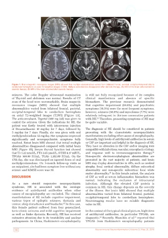
Figure 1: Brain magnetic resonance images (MRI) showed that multiple abnormalities varied from bilateral frontal, parietal, occipital-temporal lobe to
          cerebellum hemisphere on axial T2-weighted images (T2WI). Multiple abnormalities disappeared after steroids therapy. (A) MRI of the brain obtained before
          steroids therapy; (B) MRI of the brain obtained after steroids therapy
          seizures. The color  Doppler ultrasound examination   is still not fully recognized because of its complex
          of Thyroid and abdomen was normal. Results of CT    clinical  manifestions  and  absence of  specific
          scan of the head were unremarkable. Brain magnetic   biomarkers. The previous research demonstrated
          resonance images (MRI) showed  that multiple        that cognitive impairment (84.6%) and psychiatric
          abnormalities varied from bilateral frontal, parietal,   symptoms (38.5%) were the most frequent symptoms,
          occipital-temporal  lobe to cerebellum  hemisphere   however, seizures (30.8%) and myoclonus (7.7%) were
          on axial T2-weighted images (T2WI) [Figure 1A].     relatively infrequent in thirteen consecutive patients
          The anticonvulsant, Tegretol (100 mg bid) was given to   with HE.  Therefore, presenting symptoms of HE may
                                                                      [3]
          control the seizures. Given the indications for HE, the   be quite variable.
          patient  was  firstly  treated  with  intravenous  injection
          of Dexamethasone 10 mg/day for 7 days, followed by   The diagnosis of HE should be considered in patients
          5 mg/day for 7 days. Finally, she was given with oral   presenting with the characteristic neuropsychiatric
          methylprednisolone 12 mg/day. Her symptoms improved   manifestations excluding other causes of encephalopathy.
          siginificantly  and  neuropsychiatric  symptoms  fully   Generally, high levels of anti-thyroid antibodies in serum
          resolved. Retest brain MRI showed that initial multiple   or CSF are important and helpful in the diagnosis of HE.
          abnormalities disappeared compared with initial brain   They have no alteration in the CSF and/or imaging tests
          MRI [Figure 1B]. Serum thyroid function test showed   compatible with infectious, vascular, or neoplastic etiology,
                                                                                                             [4]
          that T3 1.22 nmol/L, FT3 2.65 pmol/L, S-TSH 4.17 mIU/L,   and response well to immunosuppressive therapy.
          TPOAb  369.80  IU/mL,  TGAb  1,102.00  IU/mL.  On  the   Non-specific electroencephalogram abnormalities are
          17th day, she was discharged on tapered doses of oral   presented  in  the  vast  majority  of  patients,  and  brain
          methylprednisolone. On 3-month follow-up visits as   MRI may display abnormalities in 49%, such as cerebral
          an outpatient, she had been symptom free without any   atrophy, focal cortical abnormality, diffuse subcortical
          seizure and MMSE score was 30.                      abnormality and non-specific subcortical focal white
                                                              matter abnormality.  In this female patient, the analysis
                                                                               [5]
          DISCUSSION                                          of CSF as well as serum inflammation biomarkers was
                                                              normal,  indicating  the  exclusion  of  intracranial
          As a rare steroid responsive neuropsychiatric       infection. Although the elevated CSF protein is
          syndrome, HE is  associated  with  the serologic    common in HE, this change depends on the severity
          evidence of anti-thyroid antibodies when other      of the illness. Her brain MRI showed  that multiple
          causes of encephalopathy are excluded. The clinical   abnormalities varied from bilateral frontal, parietal,
          manifestations of HE include cognitive impairment,   occipital-temporal lobe to cerebellum hemisphere.
          various  types  of epileptic seizures,  dystaxia and   Neuroimaging results have no reliable diagnostic
          tremor, sleep disturbance and headache.  In this case,   value in HE.
                                              [2]
          this female patient suffered from four generalized,
          tonic-clonic seizures and mild cognitive impairment   In the context of the typical clinical picture, high titres
          as well as limbs dystaxia. Recently, HE has received   of  antithyroid  antibodies,  in  particular TPOAb,  are
          extensive attention due to its treatability and unclear   diagnostic.  Recently, Blanchin et al.  reported that
                                                                                                [7]
                                                                        [6]
          pathogenesis. In China, Hashimoto’s encephalopathy   TPOAb from Hashimoto’s encephalopathy patients
           118                                                      Neuroimmunol Neuroinflammation | Volume 3 | May 20, 2016