
Page 130 - Read Online
P. 130
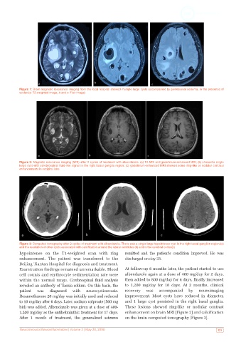
Figure 1: Brain magnetic resonance imaging from the local hospital showed multiple large cysts accompanied by perilesional oedema, in the presence of
           scolex (a: T2-weighted image; b and c: Flair image)


           Figure 2: Magnetic resonance imaging (MRI) after 2 cycles of treatment with albendazole. (a) T2-MRI and gadolinium-enhanced MRI; (b) showed a single
           large cyst with cerebrospinal fluid-like signal in the right basal ganglia region; (c) gadolinium-enhanced MRI showed some ring-like or nodular contrast
           enhancements in occipital lobe


           Figure 3: Computed tomography after 2 cycles of treatment with albendazole. There was a single large hypointense cyst in the right basal ganglion region (a)
           and the resolution of other cysts associated with calcification around the lateral ventricles (b) and in the cerebral cortex(c)
           hypointense on the T1-weighted scan with ring       remitted and the patient’s condition improved. He was
           enhancement. The patient was transferred to the     discharged on day 23.
           Beijing Tiantan Hospital for diagnosis and treatment.
           Examination findings remained unremarkable. Blood   At follow-up 6 months later, the patient started to use
           cell counts and erythrocyte sedimentation rate were   albendazole again at a dose of 400 mg/day for 2 days,
           within the normal range. Cerebrospinal fluid analysis   then added to 800 mg/day for 4 days, finally increased
           revealed an antibody of Taenia solium. On this basis, the   to 1,200 mg/day for 10 days. At 2 months, clinical
           patient  was   diagnosed  with   neurocysticercosis.  recovery  was  accompanied   by   neuroimaging
           Dexamethasone 20 mg/day was initially used and reduced   improvement. Most cysts have reduced in diameter,
           to 10 mg/day after 6 days. Later, sodium valproate (500 mg   and 1 large cyst persisted in the right basal ganglia.
           bid) was added. Albendazole was given at a dose of 400-  These lesions showed ring-like or nodular contrast
           1,200 mg/day as the antihelminthic treatment for 17 days.   enhancement on brain MRI [Figure 2] and calcification
           After 1 month of treatment, the generalized seizures   on the brain computed tomography [Figure 3].


           Neuroimmunol Neuroinflammation | Volume 3 | May 20, 2016                                          121