
Page 131 - Read Online
P. 131
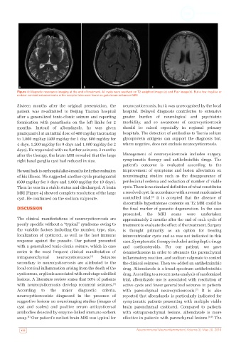
Figure 4: Magnetic resonance imaging at the end of treatment. All cysts were resolved on T2-weighted image (a) and Flair image(b). But a few ring-like or
          nodular contrast enhancements in the occipital lobe were found on gadolinium-enhanced MRI

          Sixteen months after the original presentation, the   neurocysticercosis, but it was unrecognised by the local
          patient was re-admitted to Beijing Tiantan hospital   hospital. Delayed diagnosis contributes to extensive
          after a generalized tonic-clonic seizure and reporting   greater burden of neurological and psychiatric
          formication with parasthesia on the left limbs for 2   morbidity, and so awareness of neurocysticercosis
          months. Instead of albendazole, he was given        should be raised especially in regional primary
          praziquantel at an initial dose of 400 mg/day increasing   hospitals. The detection of antibodies to Taenia solium
          to 1,800 mg/day (400 mg/day for 1 day, 600 mg/day for   glycoprotein antigens can support the diagnosis but,
          4 days, 1,200 mg/day for 9 days and 1,800 mg/day for 2   where negative, does not exclude neurocysticercosis.
          days). He responded with no further seizures. 2 months
          after the therapy, the brain MRI revealed that the large   Management of neurocysticercosis includes surgery,
          right basal ganglia cyst had reduced in size.       symptomatic therapy and antihelminthic drugs. The
                                                              patient’s outcome is evaluated according to the
          He went back to our hospital after 4 months for further evaluation   improvement of symptoms and lesion alleviation on
          of his illness. We suggested another cycle praziquantel   neuroimaging studies such as the disappearance of
          (600 mg/day for 3 days and 1,800 mg/day for 10 days).   perilesional oedema and reduction of number of viable
          Then he was in a stable status and discharged. A brain   cysts. There is no standard definition of what constitutes
          MRI [Figure 4] showed complete resolution of the large   a resolved cyst. In accordance with a recent randomised
                                                                            [6]
          cyst. He continued on the sodium valproate.         controlled  trial,   it  is  accepted  that  the  absence  of
                                                              discernible hyperintense contents on T2 MRI could be
          DISCUSSION                                          the final marker of parasite degeneration. In the case
                                                              presented,  the  MRI    scans  were   undertaken
          The clinical manifestations of neurocysticercosis are   approximately 2 months after the end of each cycle of
          poorly specific without a “typical” syndrome owing to   treatment to evaluate the effect of the treatment. Surgery
          the variable factors including the number, type, size,   is thought primarily as an option for treating
          localization of cysticerci, as well as the host immune   intraventricular cysts and so was not indicated in this
          response against the parasite. Our patient presented   case. Symptomatic therapy included antiepileptic drugs
          with a generalized tonic-clonic seizure, which in case   and corticosteroids. For our patient, we gave
          series  is  the  most  frequent  clinical  manifestation  of   dexamethasone in order to attenuate the parenchymal
          intraparenchymal   neurocysticercosis. [3]  Seizures  inflammatory reaction, and sodium valproate to control
          secondary to neurocysticercosis are attributed  to the   the clinical seizures. Then we added an antihelminthic
          local cortical inflammation arising from the death of the   drug. Albendazole is a broad-spectrum antihelminthic
          cysticercus, or gliosis associated with end-stage calcified   drug. According to a recent meta-analysis of randomized
          lesions. A literature review states that 50% of patients   trial, albendazole use is associated with resolution of
          with neurocysticercosis develop recurrent seizures.    active cysts and fewer generalized seizures in patients
                                                         [4]
          According  to   the   major   diagnostic  criteria,  with parenchymal neurocysticercosis.  It is also
                                                                                                  [7]
          neurocysticercosisis diagnosed in the presence of   reported that albendazole is particularly indicated for
          suggestive lesions on neuroimaging studies (images of   symptomatic patients presenting with multiple viable
          cyst and scolex) and positive serum anticysticercal   brain parenchymal cysticerci. Compared to patients
          antibodies detected by enzyme-linked immuno sorbent   with extraparenchymal lesions, albendazole is more
          assay.  Our patient’s earliest brain MRI was typical for   effective in patients with parenchymal lesions. [8,9]  The
               [5]

           122                                                      Neuroimmunol Neuroinflammation | Volume 3 | May 20, 2016