
Page 134 - Read Online
P. 134
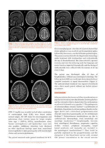
Figure 3: Sagittal fluid-attenuated inversion recovery (FLAIR) (A, B), axial
           Figure 1: Sagittal fluid-attenuated inversion recovery (FLAIR) (A, B), axial   FLAIR (C, D) and T2-weighted images (E, F) one month later, showed near
           FLAIR (C, D) and T2-weighted images (E, F) revealed multiple focal cortical   total remission of the lesions.
           areas of high signal intensity fronto-parietal and occipital (arrows) and
           “hyper-intense vein” compatible with slow flow in isolated cortical vein (curved
           arrow).                                             Electroencephalogram  after this 48 h-period showed that
                                                               status epilepticus was resolved and demonstrated spikes
                                                               followed by slow waves, over the left fronto-parietal region.
                                                               The patient was transferred to the department of neurology
                                                               under treatment with levetiracetam and corticosteroids
                                                               (40 mg of dexamethasone). She demonstrated a gradual
                                                               recovery and over the following week her language and
                                                               motor function improved dramatically and the dosing of
                                                               corticosteroids were reduced with decrements of 10 mg
                                                               every 3 days.

                                                               The patient was discharged, after 25 days of
                                                               hospitalization, without any neurological semiology. The
                                                               follow up brain MRI one month later demonstrated almost
                                                               complete remission of signal abnormalities [Figure 3].
                                                               The anticonvulsant treatment was tapered of (redcued)
                                                               over a three month period without any further seizure
                                                               occurrence.

                                                               DISCUSSION

                                                               Scleromyxedema also known as lichen myxedematosus or
                                                               papular mucinosis is a rare skin disorder involving the face
                                                               and the extremities that is characterized by erythematous
                                                               or yellowish lichenoid waxy papules. [1,2]  The pathogenesis
           Figure 2: Axial T1-weighted images before (A, B) and after intravenous              [5]
           gadolinium administration (C, D) showed cortical areas with low signal   of scleromyxedema remains unclear.  A proliferation of
           intensity (arrows) without abnormal enhancement.    fibroblasts, deposition of acid mucopolysaccharides in the
           (TPR 77 mg/dL) was recorded and all other constituents   upper dermis and a monoclonal paraproteinemia-most
           of cerebrospinal fluid (CSF) were found within      often IgG monoclonal gammopathy are the predominant
           normal ranges. All CSF stains for microorganisms and   findings. [2,5]  Extracutaneous manifestations are due to
           polymerase chain reaction assays for herpes simplex   cardiovascular, rheumatologic, renal, hematologic and
           virus type 1 (HSV-1), HSV-2, varicella-zoster virus,   neurologic involvement. [1-4]  Encephalopathy, transient
           human  cytomegalovirus  and  Epstein-Barr  virus  were   focal neurological disturbances, progressive cognitive
           also negative. Additional laboratory tests disclosed   decline,  seizures,  peripheral  neuropathy,  carpal
           no other significant findings apart from monoclonal   tunnel  syndrome and myopathy are included in
           immunoglobulin G (IgG) gammapathy with lambda light   the neurological manifestations. [2,4]  They could be
           chains and excessive proteinuria.                   partially explained by the  paraproteinemia and the
                                                               disruption of the blood-brain barrier with  elevated
           The patient remained under general anesthesia for 48 h.   CSF protein concentration in the central nervous


           Neuroimmunol Neuroinflammation | Volume 3 | May 25, 2016                                          125