
Page 132 - Read Online
P. 132
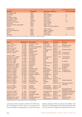
Table 1: Immunohistochemical evaluation result for primary malignant ganglionic paraganglioma
           Antibody                          Distributor         Abbreviation (dilution)        Immunoreactivity
           Neurofilament MAb                 DAKO                NF (1:400)                     -
           Neural CAM                        DAKO                CD56 (undiluted)               +++
           β-tubulin                         SIGMA               βTUB (1:5000)                  F++
           Synaptophysin MAb                 DAKO                SYN (1:25)                     ++
           Chromogranin A MAb                DAKO                CHRG (1:100)                   ++
           Calretinin Poly Ab                Invitrogen          CAL (1:200)                    F+
           Neuron‑specific enolase           DAKO                NSE (undiluted)                +, ++
           S100 protein                      DACO                S100 (1:4000)                  -
           Epithelial membrane antigen MAb   DACO                EMA (1:100)                    -
           Keratins                          DACO                CAM5.2, AE1/AE3 (undiluted)    -
                                                                                                ++ sustentacular c.
           GFAP Poly Ab                      DAKO                GFAP (1:3000)                  + sustentacular c.
           Anti-human melanosome             DAKO                HMB 45 (undiluted)             -
           Desmin                            DACO                DSM (undiluted)                -
           p53                               DAKO                p53 (1:200)                    +
           Ki-67 MAb                         DAKO                Ki-67 (1:1000)                 +++
           GFAP: glial fibrillary acidic protein; CAM: cell adhesion molecule; MAb: monoclonal antibody


           Table 2: Literature review for intracranial supratentorial primary paraganglioma
           Author              Age (gender)  Presentation       Location      Treatment    Outcome
           Kruse, 1960 [10]      68, male  Behavioral changes   Middle fossa  Resection    Improved
           Smith et al., 1966* [11]  17, male  Headache         Pineal region  Resection   Moderate disability
           Chytil, 1967 [12]     46, male  Visual loss, hypopituitarism  Sellar/suprasellar  Resection + RT  No progression
           Bilbao et al., 1978 [13]  37, male  Delayed growth   Sellar        Resection    -
           Ho et al., 1982 [14]  65, male  Diplopia             Cavernous sinus  Resection  Moderate disability
           Prabhakar et al., 1984 [15]  7, female  Ophthalmoplegia  Parasellar  Resection + RT  -
           Steel et al., 1993 [16]  44, female  Headache        Sellar        Resection + RT  No progression
                                41, female  Headache, ptosis    Sellar        Resection + RT  No progression
           Flint et al., 1993 [17]  17, female  Visual defect   Sellar        Resection    -
           Scheithauer et al., 1996 [18]  14, male  Visual defect  Sellar/parasellar  Resection + RT  Left hemiparesis
           Nishitani et al., 1996* [19]  41, female  Amenorrhea  Parasellar   Resection    Good recovery
           Noble et al., 1997 [8]  71, male  Visual defect      Sellar        Resection    -
           Mokry et al., 1998 [9]  76, female  Visual defect    Sellar        Resection    Unchanged
           Caro et al., 1998 [20]  84, male  Memory loss        Sellar/suprasellar  Resection  -
           Sambaziotis et al., 1999 [21]  54, male  Visual defect  Sellar     Resection    No progression
           Yamauchi et al., 1999 [22]  56, female  Headache     Frontal fossa  Resection   No progression
           Reithmeier et al., 2000 [23]  42, male  Seizure      Insula        Resection    Hemiparesis
           Laquis et al., 2001 [24]  15, female  Occulomotor palsy  Middle fossa  Resection + RT  Improved
           Salame et al., 2001 [25]  48, female  Oligomenorrhea  Sellar/parasellar  Resection  No progression
           Hertel et al., 2003 [26]  51, female  Facial paresis  Middle fossa  Resection + RT  Occulomotor palsy
           Yokoo et al., 2003 [27]  52, female  Behavioral changes  Suprasellar  Resection  -
           Arkha et al., 2003 [28]  58, female  Endocrine dysfunction  Sellar/parasellar  Resection  -
           Riopel et al., 2004 [29]  66, male  Diplopia         Parasellar    Biopsy       -
           Naggara et al., 2005 [30]  47, male  Visual defect   Suprasellar   Resection    -
           Zorlu et al., 2005 [31]  37, male  Visual defect     Sellar/suprasellar  Resection + RT  -
           Boari et al., 2006 [32]  52, male  Brain ischemia    Sellar        Resection    Pituitary dysfunction
           Peltier et al., 2007 [33]  51, female  Occulomotor palsy  Parasellar  Resection  -
           Sinha et al., 2008 [34]  18, male  Visual defect     Sellar        Resection + RT  Skull, scalp and femur
                                                                                           metastasis
           Yoo et al., 2008 [35]  21, female  Headache          Temporal lobe  Resection + RT  -
           Ozüm et al., 2008 [36]  70, male  Headache           Sellar/parasellar  Resection + RT  -
           Lu et al., 2009 [5]   81, male  Visual change        Sellar/suprasellar  Resection  Died 4 months after
                                                                                           (esophageal cancer)
           Haresh et al., 2009 [37]  17, male  Visual change    Sellar/suprasellar  Resection + RT  Skull and femur metastasis
           Thakar et al., 2011 [38]  40, male  Visual defect    Frontal lobe  Resection    Recurrence (6 months)
           Prajsnar et al., 2011 [39]  53, female  Trigeminal neuralgia  Meckel’s cave  Resection  Recurrence (2 years)
           Albert et al., 2011 [40]  63, male  Proptosis        Sellar/parasellar  Resection + RT  Improved
           Nascimento et al., 2012 [41]  33, female  Endocrine dysfunction  Sellar  Resection  Diabetes insipidus
           Chaudhry et al., 2013 [42]  44, male  Visual defect  Sellar/suprasellar  Resection  No recurrence
           *Found in Yamauchi et al. review of literature, 1999. RT: radiation therapy
           component makes it unique and the first of this type.   primary malignant forms are known. In addition, the
           The  radiological features  were not conclusive  for   tumor was not functional, and it did not associate with
           primary malignant paraganglioma as only extracranial   any hemodynamic changes intra-operatively to alert for


            124                                              Neuroimmunol Neuroinflammation | Volume 2 | Issue 2 | April 15, 2015