
Page 127 - Read Online
P. 127
a b
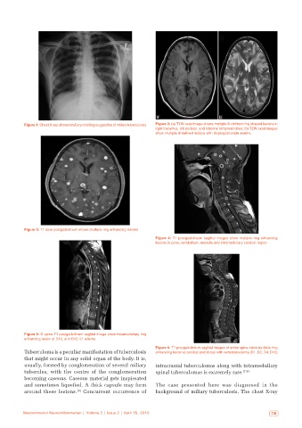
Figure 1: Chest X-ray showed miliary mottling suggestive of miliary tuberculosis Figure 2: (a) T1W axial image shows multiple ill‑defined ring shaped lesions in
right thalamus, left parietal, and bilateral temporal lobes; (b) T2W axial images
show multiple ill‑defined lesions with disproportionate edema
Figure 3: T1 axial postgadolinium shows multiple ring enhancing lesions
Figure 4: T1 postgadolinium sagittal images show multiple ring enhancing
lesions in pons, cerebellum, medulla and intramedullary cervical region
Figure 5: D spine T1 postgadolinium sagittal image show intramedullary ring
enhancing lesion at D10, and D12, L1 edema
Figure 6: T1 postgadolinium sagittal images of entire spine show multiple ring
Tuberculoma is a peculiar manifestation of tuberculosis enhancing lesion at cervical and dorsal with vertebral edema (D1, D2, D4, D12)
that might occur in any solid organ of the body. It is,
usually, formed by conglomeration of several miliary intracranial tuberculomas along with intramedullary
tubercles, with the centre of the conglomeration spinal tuberculomas is extremely rare. [7,8]
becoming caseous. Caseous material gets inspissated
and sometimes liquefied. A thick capsule may form The case presented here was diagnosed in the
[6]
around these lesions. Concurrent occurrence of background of miliary tuberculosis. The chest X-ray
Neuroimmunol Neuroinflammation | Volume 2 | Issue 2 | April 15, 2015 119