
Page 131 - Read Online
P. 131
a b
Figure 3: Brain magnetic resonance imaging after 6 weeks from surgery depicts
a tumor bed cavity with complete resection. A slight gadolinium uptake was seen
within the tumor cavity which represents remnant of fibrillary surgi‑cells packing
(ethicon) from the second surgery c d
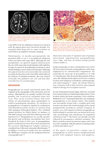
Figure 4: (a) Large neoplastic ganglionic cell with prominent nucleoli and
scale (KPS) of 30, the radiation treatment was deferred cytoplasmic vacuolation HE; (b) strong chromogranin immunoreactivity in
epitheliod tumor cells; (c) cytoplasmic synaptophysin immunoreactivity in a large
until the patient gains more functional strength. She ganglionic cell and the processes of the adjacent tumor cells; (d) glial fibrillary
remained stable along with no tumor recurrence on acidic protein immunoreaction accentuated at the periphery of the tumor nest
serial follow-up magnetic resonance imaging. characteristically seen in sustentacular paraganglioma cells
Unfortunately, 12 months postoperatively she There have been only 37 reported cases of primary
presented with a 5 cm × 5 cm ipsilateral enhancing intracranial supratentorial paragangliomas
lesion associated with mass effect. Although she was since 1960, and they all harbor benign growth
asymptomatic, we opted for surgical resection given features [Table 2].
the size of the mass that would interfere with radiation
therapy. As expected, the pathological work up revealed In this manuscript, we have excluded from our review
similar findings to previously resected tumor. Although all intracranial metastatic paragangliomas and lesions
her surgical resection went uneventful, she continued originated in the infratentorial compartment. We
to decline her functions with lower KPS, which affected found that the mean age of presentation is 47 with
the delivery of radiation treatment. She was referred 1:1.3 female:male ratio. In around three-quarter of these
eventually into palliative care unit and died after cases, the sellar/parasellar region was the most common
2 months from her second surgery. location with symptoms of headache, opthalmoplegia,
and endocrinopathy. [43] Again, the benign behavior
DISCUSSION was a character of all of them with few cases required
radiation therapy for incomplete resection.
Paragangliomas are neural crest-derived tumors that
originate in the paraganglia of the autonomic nervous In term of intraparenchymal origin, only three cases had
system. Although they are usually considered benign been reported and were harboring benign pattern. Their
growths, but occasionally malignant forms may location within the brain represents a rare condition.
occur. They can be functional lesions with active Reithmeier et al. had reported the first case of primary
[23]
release of catecholamines upon manipulation to paraganglioma in the insular region. This patient
result in hemodynamic alterations. The incidence of was successfully treated with a complete gross total
paraganglioma is frequently reported in combination resection, but dense left sided hemiplegia had occurred
with pheochromocytoma owing to its rareness along postoperatively secondary to cerebral vasospasm. In
with analogous histological features. The combined another case, a left temporal melanotic paraganglioma
annual incidence in United States, for instance, is had developed in a patient who was treated previously
500-1600 cases per year with 50% of them present with with chemotherapy and radiotherapy for langerhans
hypertension. Paragangliomas are usually sporadic cell histiocytosis. [35] The lesion was partially resected
[3]
but genetic and syndromic associations have been followed by postoperative radiation treatment. The
described. In about 27-32% cases of paraganglioma, third case had occurred in premotor region, and it was
genetic mutations have been discovered among which resected completely with the patient remaining stable
the succinate dehydrogenase complex mutation is at short-term follow-up. [38]
considered one of the most commonly affected genes. [4,5]
In addition, several recent works have also correlated In this report, we presented a unique case of
the SDHB gene mutation and the malignant behavior paraganglioma in which primary intraparenchymal
of paragangliomas. [6-9] growth with malignant features and ganglionic
Neuroimmunol Neuroinflammation | Volume 2 | Issue 2 | April 15, 2015 123