
Page 128 - Read Online
P. 128
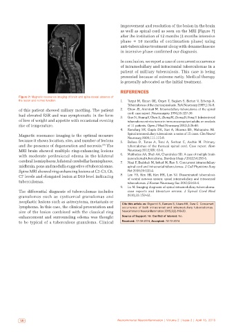
improvement and resolution of the lesion in the brain
                                                              as well as spinal cord as seen on the MRI [Figure 7]
                                                              after the institution of 12 months (2 months intensive
                                                              phase  +  10 months of continuation phase) using
                                                              anti-tuberculous treatment along with dexamethasone
                                                              in intensive phase confirmed our diagnosis.

                                                              In conclusion, we report a case of concurrent occurrence
                                                              of intramedullary and intracranial tuberculomas in a
                                                              patient of military tuberculosis. This case is being
                                                              presented because of extreme rarity. Medical therapy
                                                              is generally advocated as the initial treatment.

                                                              REFERENCES
           Figure 7: Magnetic resonance imaging of brain and spine reveal absence of
           the lesion and normal function                     1.   Turgut M, Ozcan OE, Ozgen T, Saglam S, Bertan V, Erbengi A.
                                                                  Tuberculomas of the craniospinal axis. Turk Neurosurg 1989;1:34‑8.
           of this patient showed miliary mottling. The patient   2.   Citow JS, Ammirati M. Intramedullary tuberculoma of the spinal
           had elevated ESR and was symptomatic in the form   3.   cord: case report. Neurosurgery 1994;35:327‑30.
                                                                  Guo N, Huang F, Chen X, Zheng M, Zhong D, Feng Y. Infratentorial
           of loss of weight and appetite with occasional evening   tuberculoma mimics tumor in immunocompetent adults: an analysis
           rise of temperature.                                   of 11 patients. Open J Mod Neurosurg 2013;3:36‑40.
                                                              4.   Ramdurg SR, Gupta DK, Suri A, Sharma BS, Mahapatra AK.
           Magnetic resonance imaging is the optimal measure      Spinal intramedullary tuberculosis: a series of 15 cases. Clin Neurol
                                                                  Neurosurg 2009;111:115‑8.
           because it shows location, size, and number of lesions   5.   Balasa D, Tunas A, Terzi A, Serban C, Aschie M. Primary
           and the presence of degeneration and necrosis.  The    tuberculomas of the thoracal spinal cord. Case report.  Rom
                                                     [9]
           MRI brain showed multiple ring-enhancing lesions       Neurosurg 2012;XIX: 63‑6.
           with  moderate  perilesional  edema  in  the  bilateral   6.   Mukherjee AA, Shah AA, Chandrakar SD. A case of multiple brain
                                                                  parenchymal tuberculoma. Bombay Hosp J 2012;54:293‑6.
           cerebral hemispheres, bilateral cerebellar hemispheres,   7.   Niazi F, Badshah M, Irshad M, Rao S. Concurrent intramedullary
           midbrain, pons and medulla suggestive of tuberculomas.   spinal cord and intracranial tuberculomas. J Coll Physicians Surg
           Spine MRI showed ring enhancing lesions at C2-C3, C6,   Pak 2010;20:132‑4.
           C7 levels and elongated lesion at D10 level indicating   8.   Lim YS, Kim SB, Kim MK, Lim YJ. Disseminated tuberculosis
                                                                  of central nervous system: spinal intramedullary and intracranial
           tuberculomas.                                          tuberculomas. J Korean Neurosurg Soc 2013;54:61‑4.
                                                              9.   Lu M. Imaging diagnosis of spinal intramedullary tuberculoma:
           The differential diagnosis of tuberculomas includes    case reports and literature review.  J  Spinal Cord Med
           granulomas such as cysticercal granulomas and          2010;33:159‑62.
           neoplastic lesions such as astrocytoma, metastasis or   Cite this article as: Diguvinti S, Damam S, Ubara KK, Dara C. Concurrent
           lymphoma. In this case, the clinical presentation and   occurrence  of  both  intracranial  and  intramedullary  tuberculomas.
           size of the lesion combined with the classical ring   Neuroimmunol Neuroinflammation 2015;2(2):118-20.
           enhancement and surrounding edema was thought       Source of Support: Nil. Conflict of Interest: No.
           to be typical of a tuberculous granuloma. Clinical   Received: 17-09-2014; Accepted: 14-10-2014


            120                                              Neuroimmunol Neuroinflammation | Volume 2 | Issue 2 | April 15, 2015