
Page 98 - Read Online
P. 98
hyperintensities affecting the posterior aspects of the
brain, namely the occipital and parietal lobes. It is
now known that this description is more of a general
rule, and those asymmetric images can be seen, and
can involve the deep grey matter as well as the frontal
and temporal lobes. The advent of diffusion weighted
imaging helped clarify that the MRI changes were
not due to ischemia or cytotoxic edema, but due to
vasogenic edema. [7]
In our case, as per diagnostic criteria for SLE, more
[8]
than four well documented features were present, that
is, history of arthralgias, photosensitivity, polyserositis,
renal impairment and nervous system involvement.
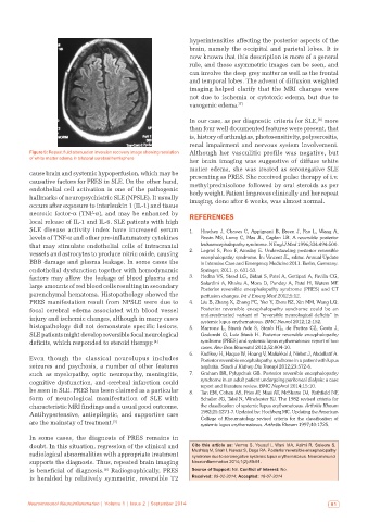
Figure 5: Repeat fluid attenuation inversion recovery image showing resolution Although her vasculitic profile was negative, but
of white matter edema in bilateral cerebral hemisphere
her brain imaging was suggestive of diffuse white
matter edema, she was treated as seronegative SLE
cause brain and systemic hypoperfusion, which may be presenting as PRES. She received pulse therapy of i.v.
causative factors for PRES in SLE. On the other hand, methylprednisolone followed by oral steroids as per
endothelial cell activation is one of the pathogenic body weight. Patient improves clinically and her repeat
hallmarks of neuropsychiatric SLE (NPSLE). It usually imaging, done after 6 weeks, was almost normal.
occurs after exposure to interleukin 1 (IL-1) and tissue
necrotic factor-α (TNF-α), and may be enhanced by REFERENCES
local release of IL-1 and IL-6. SLE patients with high
SLE disease activity index have increased serum 1. Hinchey J, Chaves C, Appignani B, Breen J, Pao L, Wang A,
levels of TNF-α and other pro-inflammatory cytokines Pessin MS, Lamy C, Mas JL, Caplan LR. A reversible posterior
that may stimulate endothelial cells of intracranial leukoencephalopathy syndrome. N Engl J Med 1996;334:494-500.
vessels and astrocytes to produce nitric oxide, causing 2. Legriel S, Pico F, Azoulay E. Understanding posterior reversible
encephalopathy syndrome. In: Vincent JL, editor. Annual Update
BBB damage and plasma leakage. In some cases the in Intensive Care and Emergency Medicine 2011. Berlin, Germany:
endothelial dysfunction together with hemodynamic Springer; 2011. p. 631-53.
factors may allow the leakage of blood plasma and 3. Hedna VS, Stead LG, Bidari S, Patel A, Gottipati A, Favilla CG,
large amounts of red blood cells resulting in secondary Salardini A, Khaku A, Mora D, Pandey A, Patel H, Waters MF.
Posterior reversible encephalopathy syndrome (PRES) and CT
parenchymal hematoma. Histopathology showed the perfusion changes. Int J Emerg Med 2012;5:12.
PRES manifestation result from NPSLE were due to 4. Liu B, Zhang X, Zhang FC, Yao Y, Zhou RZ, Xin MM, Wang LQ.
focal cerebral edema associated with blood vessel Posterior reversible encephalopathy syndrome could be an
injury and ischemic changes, although in many cases underestimated variant of “reversible neurological deficits” in
systemic lupus erythematosus. BMC Neurol 2012;12:152.
histopathology did not demonstrate specific lesions. 5. Marrone L, Streck Ade S, Staub HL, de Freitas CZ, Costa J,
SLE patients might develop reversible focal neurological Gadonski G, Luiz Staub H. Posterior reversible encephalopathy
deficits, which responded to steroid therapy. [4] syndrome (PRES) and systemic lupus erythematosus: report of two
cases. Rev Bras Reumatol 2012;52:804-10.
6. Kadikoy H, Haque W, Hoang V, Maliakkal J, Nisbet J, Abdellatif A.
Even though the classical neurolupus includes Posterior reversible encephalopathy syndrome in a patient with lupus
seizures and psychosis, a number of other features nephritis. Saudi J Kidney Dis Transpl 2012;23:572-6.
such as myelopathy, optic neuropathy, meningitis, 7. Graham BR, Pylypchuk GB. Posterior reversible encephalopathy
cognitive dysfunction, and cerebral infarction could syndrome in an adult patient undergoing peritoneal dialysis: a case
report and literature review. BMC Nephrol 2014;15:10.
be seen in SLE. PRES has been claimed as a particular 8. Tan EM, Cohen AS, Fries JF, Masi AT, McShane DJ, Rothfield NF,
form of neurological manifestation of SLE with Schaller JG, Talal N, Winchester RJ. The 1982 revised criteria for
characteristic MRI findings and a usual good outcome. the classification of systemic lupus erythematosus. Arthritis Rheum
Antihypertensive, antiepileptic, and supportive care 1982;25:1271-7. Updated by: Hochberg MC. Updating the American
College of Rheumatology revised criteria for the classification of
are the mainstay of treatment. [5] systemic lupus erythematosus. Arthritis Rheum 1997;40:1725.
In some cases, the diagnosis of PRES remains in
doubt. In this situation, regression of the clinical and Cite this article as: Verma S, Yousuf I, Wani MA, Asimi R, Saleem S,
radiological abnormalities with appropriate treatment Mushtaq M, Shah I, Nawaz S, Daga RA. Posterior reversible encephalopathy
syndrome due to seronegative systemic lupus erythematosus. Neuroimmunol
supports the diagnosis. Thus, repeated brain imaging Neuroinflammation 2014;1(2):89-91.
is beneficial of diagnosis. Radiographically, PRES Source of Support: Nil. Conflict of Interest: No.
[6]
is heralded by relatively symmetric, reversible T2 Received: 09-02-2014; Accepted: 18-07-2014
Neuroimmunol Neuroinflammation | Volume 1 | Issue 2 | September 2014 91