
Page 103 - Read Online
P. 103
in tuberculous meningitis occurs in 15-57% of patients
especially in advanced stage and severe illness and
are usually multiple, bilateral and located in the
basal ganglia, especially the tubercular zone, which
comprises of the caudate, anterior thalamus, anterior
limb and genu of the internal capsule. Cortical
a b stroke can also occur due to the involvement of
the proximal portion of the middle, anterior and
posterior cerebral arteries, as well as the supraclinoid
portion of the internal carotid and basilar arteries.,
[1]
While pathological changes suggestive of intracranial
vasculitis are common in tuberculosis even without
corresponding clinical features, to our knowledge,
c d this is the first reported case of malignant MCA
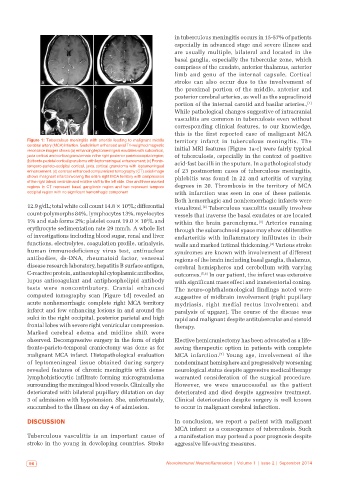
Figure 1: Tuberculous meningitis with arteritis leading to malignant middle territory infarct in tuberculous meningitis. The
cerebral artery (MCA) infartion. Gadolinium enhanced axial T1‑weighted magnetic
resonance images shows (a) enhancing leptomeningeal exudates with subcortical, initial MRI features [Figure 1a-c] were fairly typical
juxta cortical and cortical granulomata in the right posterior parietooccipital region; of tuberculosis, especially in the context of positive
(b) fronto‑parietal cortical granuloma with leptomeningeal enhancement; (c) Fronto‑ acid-fast bacilli in the sputum. In a pathological study
temporo‑parieto‑occipital cortical, juxta cortical granuloma with leptomeningeal
enhancement. (d) contrast enhanced computerized tomography (CT) axial image of 23 postmortem cases of tuberculous meningitis,
shows malignant infarct involving the entire right MCA territory with compression phlebitis was found in 22 and arteritis of varying
of the right lateral ventricle and midline shift to the left side. One and three marked
regions in CT represent basal ganglionic region and two represent temporo degrees in 20. Thrombosis in the territory of MCA
occipital region with no significant hemorrhagic component with infarction was seen in one of these patients.
Both hemorrhagic and nonhemorrhagic infarcts were
12.9 g/dL; total white cell count 14.8 × 10 /L; differential visualized. Tuberculous vasculitis usually involves
9
[2]
count-polymorphs 84%, lymphocytes 13%, myelocytes vessels that traverse the basal exudates or are located
1% and stab forms 2%; platelet count 19.0 × 10 /L and within the brain parenchyma. Arteries running
9
[3]
erythrocyte sedimentation rate 29 mm/h. A whole list through the subarachnoid space may show obliterative
of investigations including blood sugar, renal and liver endarteritis with inflammatory infiltrates in their
functions, electrolytes, coagulation profile, urinalysis, walls and marked intimal thickening. Various stroke
[4]
human immunodeficiency virus test, antinuclear syndromes are known with involvement of different
antibodies, ds-DNA, rheumatoid factor, venereal regions of the brain including basal ganglia, thalamus,
disease research laboratory, hepatitis B surface antigen, cerebral hemispheres and cerebellum with varying
C-reactive protein, antineutophil cytoplasmic antibodies, outcomes. [5,6] In our patient, the infarct was extensive
lupus anticoagulant and antiphospholipid antibody with significant mass effect and transtentorial coning.
tests were noncontributory. Cranial enhanced The neuro-ophthalomological findings noted were
computed tomography scan [Figure 1d] revealed an suggestive of midbrain involvement (right pupillary
acute nonhemorrhagic complete right MCA territory mydriasis, right medial rectus involvement and
infarct and few enhancing lesions in and around the paralysis of upgaze). The course of the disease was
sulci in the right occipital, posterior parietal and high rapid and malignant despite antitubercular and steroid
frontal lobes with severe right ventricular compression. therapy.
Marked cerebral edema and midline shift were
observed. Decompressive surgery in the form of right Elective hemicraniectomy has been advocated as a life-
fronto-parieto-temporal craniectomy was done as for saving therapeutic option in patients with complete
malignant MCA infarct. Histopathological evaluation MCA infarction. Young age, involvement of the
[7]
of leptomeningeal tissue obtained during surgery nondominant hemisphere and progressively worsening
revealed features of chronic meningitis with dense neurological status despite aggressive medical therapy
lymphohistiocytic infiltrate forming microgranuloma warranted consideration of the surgical procedure.
surrounding the meningeal blood vessels. Clinically she However, we were unsuccessful as the patient
deteriorated with bilateral pupillary dilatation on day deteriorated and died despite aggressive treatment.
3 of admission with hypotension. She, unfortunately, Clinical deterioration despite surgery is well known
succumbed to the illness on day 4 of admission. to occur in malignant cerebral infarction.
DISCUSSION In conclusion, we report a patient with malignant
MCA infarct as a consequence of tuberculosis. Such
Tuberculous vasculitis is an important cause of a manifestation may portend a poor prognosis despite
stroke in the young in developing countries. Stroke aggressive life-saving measures.
96 Neuroimmunol Neuroinflammation | Volume 1 | Issue 2 | September 2014