
Page 39 - Read Online
P. 39
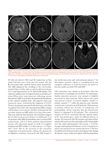
a                       b                        c                        d


           e                        f                       g                        h
          Figure 1: Axial fluid attenuated inversion recovery images of involvement in different areas: (a) Involvement in the bilateral thalamus and basal ganglia; (b) Involvement
          in the bilateral thalamus; (c) Involvement in the left prefrontal cortex; (d) Involvement in the bilateral frontal lobe, temporal lobe, insula cortex, thalamus, and basal
          ganglia; (e) Involvement in the bilateral hippocampal tail; (f) Involvement in the bilateral hippocampal head and body and uncus; (g) Involvement in the corpus callosum;
          (h) Involvement in the bilateral hippocampal head and body and midbrain

          We did not observe NCC and JE coinfection in this   rise in the late-acute and early-subacute phases. [17]  In
          study, but there was a forty-year-old woman who got   this phase, cytotoxic edema is accompanied by the
          JE associated with cerebral venous sinus thrombosis.   vasogenic collection of fluid that allows the lesion to
          The MRI displayed the swelling of the left frontal,   become visible on both T2W and DWI.
          parietal lobes and the right occipital with hypointense
          on T1 and hyperintense on T2 and FLAIR images. The   CSF parameters may change in JE patients when the
          superior sagittal sinus, left sigmoid sinus and transverse   brain tissues or meninges are involved. To summarize,
          sinus filling defect were observed, indicating venous   lumbar  puncture  pressure  was  normal  or  slightly
          thrombosis. MRV scan further demonstrated thrombosis   increased in most patients. White blood cell count
          in the superior sagittal sinus, left sigmoid sinus and   and protein content increased slightly, similar to
          transverse sinus, confirming the diagnosis of CVST.   related reports, [23,24]  while the glucose and chloride
          Twenty-six patients presented with dystonia, including   content were normal in most JE patients. This increase
          16 patients (61.54%) who demonstrated MRI changes   in protein reflects the increase of endothelial cell
          with thalamic involvement. Another 16 JE patients did   permeability, which indicates that the blood-brain
          not show lesions on MRI within 6-22 days after onset.   barrier is damaged in JE patients. The cytological
          This may indicate that thalamic abnormalities suggest   features of CSF are associated with the clinical course
          JE, but absence does not exclude it. In additional, 11 of   of JE, and performance varies in different phases of
          30 patients with MRI abnormalities also received DWI   disease. We analyzed 108 CSF samples and observed the
          examinations, and 9 of these patients demonstrated   mixed-cell reaction in the early phase of JE. Neutrophils
          lesions with abnormal signals on both T2-weighted   were the major inflammatory cells (mean = 35.67%),
          imaging and DWI within 5-12 days (mean = 5.9 days)   though neutrophil predominance is not uncommon in
          after onset. Due to the decrease in the vasculitis   other viral central nervous system infections. [25]  We also
          component and perivascular cuffing, the proportion   identified activated monocytes and plasma cells during
          of diffusion restriction decreased and ADC began to   the disease course. In the acute phase of JE, the proportion


            32                                                 Neuroimmunol Neuroinflammation | Volume 1 | Issue 1 | June 2014