
Page 40 - Read Online
P. 40
In endemic areas and epidemic season, the patients
considered as CNS infection with bilateral thalamic
involvement should be highly suspected as JE.
REFERENCES
a b 1. Solomon T. Control of Japanese encephalitis – within our grasp? N
Engl J Med 2006;355:869‑71.
2. Erlanger TE, Weiss S, Keiser J, Utzinger J, Wiedenmayer K. Past,
present, and future of Japanese encephalitis. Emerg Infect Dis
2009;15:1‑7.
3. Gao X, Nasci R, Liang G. The neglected arboviral infections in
mainland China. PLoS Negl Trop Dis 2010;4:e624.
4. Basumatary LJ, Raja D, Bhuyan D, Das M, Goswami M, Kayal AK.
Clinical and radiological spectrum of Japanese encephalitis. J Neurol
c d Sci 2013;325:15‑21.
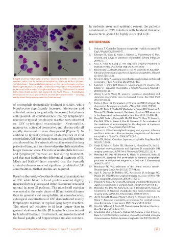
Figure 2: May–Grünwald–Giemsa staining results (×1000) of the 5. Ghosh D, Basu A. Japanese encephalitis‑a pathological and clinical
cerebral spinal fluid in Japanese encephalitis patient at different phases: perspective. PLoS Negl Trop Dis 2009;3:e437.
(a) Initial phase – Neutrophils (black arrows) are the main inflammatory cells in 6. Solomon T, Dung NM, Kneen R, Gainsborough M, Vaughn DW,
the background; (b) Acute phase – A decrease in the number of neutrophils and Khanh VT. Japanese encephalitis. J Neurol Neurosurg Psychiatry
an increase in the number of lymphocytes were noted. Furthermore, activated
monocytes (black arrows) are apparent; (c) Acute phase – Plasmocytes 2000;68:405‑15.
developed in the acute phase (black arrows); (d) Convalescence – Cytology 7. Zheng Y, Li M, Wang H, Liang G. Japanese encephalitis and
mainly shows the lymphocyte reaction (black arrows) Japanese encephalitis virus in mainland China. Rev Med Virol
2012;22:301‑22.
8. Kalita J, Misra UK. Comparison of CT scan and MRI findings in the
of neutrophils dramatically declined to 5.82%, while diagnosis of Japanese encephalitis. J Neurol Sci 2000;174:3‑8.
lymphocytes significantly increased. Monocytes and 9. Misra UK, Kalita J, Phadke RV, Wadwekar V, Boruah DK, Srivastava A,
activated monocytes gradually decreased, but plasma Maurya PK, Bhattacharyya A. Usefulness of various MRI sequences
cells peaked. At convalescence, mainly lymphocyte in the diagnosis of viral encephalitis. Acta Trop 2010;116:206‑11.
reaction or typical lymphocyte reaction were observed 10. Dung NM, Turtle L, Chong WK, Mai NT, Thao TT, Thuy TT, Kneen R,
Phu NH, Wills B, Farrar J, Das K, Solomon T. An evaluation of
on CSF cytological examination. Neutrophils, the usefulness of neuroimaging for the diagnosis of Japanese
monocytes, activated monocytes, and plasma cells all encephalitis. J Neurol 2009;256:2052‑60.
rapidly decreased or even disappeared [Figure 2]. In 11. Sawlani V. Diffusion‑weighted imaging and apparent diffusion
addition to typical cytological characteristics of viral coefficient evaluation of herpes simplex encephalitis and Japanese
encephalitis. J Neurol Sci 2009;287:221‑6.
encephalitis, CSF cytological examination of JE patients 12. Misra UK, Kalita J. Overview: Japanese encephalitis. Prog Neurobiol
also showed that the mixed-cell reaction existed for long 2010;91:108‑20.
periods of time, and we observed neutrophils existed for 13. Singh P, Kalra N, Ratho RK, Shankar S, Khandelwal N, Suri S.
longer than one week. The rates of neutrophils decrease Coexistent neurocysticercosis and Japanese B encephalitis: MR
imaging correlation. AJNR Am J Neuroradiol 2001;22:1131‑6.
and lymphocyte increase are fast during treatment, 14. Handique SK, Das RR, Barman K, Medhi N, Saharia B, Saikia P,
and this may facilitate the differential diagnosis of JE. Ahmed SA. Temporal lobe involvement in Japanese encephalitis:
Misra and Kalita [26] have reported that the 3-month problems in differential diagnosis. AJNR Am J Neuroradiol
2006;27:1027‑31.
clinical outcomes were not significantly related to CSF 15. Handique SK. Viral infections of the central nervous system.
abnormalities. Further studies are required. Neuroimaging Clin N Am 2011;21:777‑94, vii.
16. Agid R, Ducreux D, Halliday WC, Kucharczyk W, terBrugge KG,
Based on the results of routine biochemical examinations Mikulis DJ. MR diffusion‑weighted imaging in a case of West Nile
virus encephalitis. Neurology 2003;61:1821‑3.
of CSF, white blood cell and protein content slightly 17. Prakash M, Kumar S, Gupta RK. Diffusion‑weighted MR imaging in
increased while glucose and chloride content remained Japanese encephalitis. J Comput Assist Tomogr 2004;28:756‑61.
normal in most JE patients. The mixed-cell reaction 18. Handique SK, Das RR, Saharia B, Das P, Buragohain R, Saikia P.
was noted in the early phase of JE and existed longer Coinfection of Japanese encephalitis with neurocysticercosis: an
imaging study. AJNR Am J Neuroradiol 2008;29:170‑5.
than in general viral encephalitis. At convalescence, 19. Jia M, Xiong N, Huang J, Wang Y, Zhang X, Zhang Z, Cao X, Lin Z,
cytological examinations of CSF demonstrated mainly Wang T. Japanese encephalitis accompanied by cerebral venous
lymphocyte reaction or typical lymphocyte reaction. sinus thrombosis: a case report. BMC Neurol 2012;12:43.
The mixed-cell reaction in JE lasts longer than in 20. Sips GJ, Wilschut J, Smit JM. Neuroinvasive flavivirus infections.
Rev Med Virol 2012;22:69‑87.
general viral encephalitis. JE imaging is characterized 21. Ghoshal A, Das S, Ghosh S, Mishra MK, Sharma V, Koli P, Sen E,
by bilateral thalamic involvement, and involvement of Basu A. Proinflammatory mediators released by activated microglia
the basal ganglia and hippocampus are also common. induces neuronal death in Japanese encephalitis. Glia 2007;55:483‑96.
Neuroimmunol Neuroinflammation | Volume 1 | Issue 1 | June 2014 33