
Page 852 - Read Online
P. 852
Melillo et al. Mini-invasive Surg 2020;4:81 I http://dx.doi.org/10.20517/2574-1225.2020.83 Page 9 of 17
A B
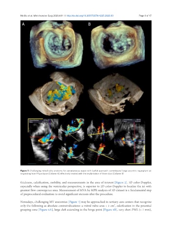
Figure 7. Challenging mitral valve anatomy for percutaneous repair with leaflet approach: commissural large eccentric regurgitant jet
originating from P3 prolapse (Column A) effectively treated with the implantation of three clips (Column B)
thickness, calcification, mobility, and measurements in the area of interest [Figure 2]. 3D color-Doppler,
especially when using the ventricular perspective, is superior to 2D color-Doppler to localize the jet with
greatest flow convergence area. Measurement of MVA by MPR analysis of 3D dataset is a fundamental step
of preprocedural evaluation to avoid significant stenosis after the procedure.
Nowadays, challenging MV anatomies [Figure 7] may be approached in tertiary care centers that recognize
2
only the following as absolute contraindications: a mitral valve area < 3 cm , calcification in the potential
grasping zone [Figure 8A], large cleft extending to the hinge point [Figure 8B], very short PML (< 7 mm),