
Page 853 - Read Online
P. 853
Page 10 of 17 Melillo et al. Mini-invasive Surg 2020;4:81 I http://dx.doi.org/10.20517/2574-1225.2020.83
A B
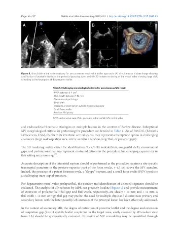
Figure 8. Unsuitable mitral valve anatomy for percutaneous repair with leaflet approach: (A) simultaneous biplane image showing
calcification of posterior leaflet in the potential grasping zone; and (B) 3D volume rendering of the mitral valve showing large cleft
extending to the hinge point of the posterior leaflet
Table 1. Challenging morphological criteria for percutaneous MV repair
MVA between 3-4 cm 2
PML length between 7-10 mm
Commissural pathology
Small cleft
Presence of calcification outside the grasping zone
Small fossa ovalis
Previous MV plasty
MVA: mitral valve area; PML: posterior mitral leaflet; MV: mitral valve
and endocarditic/rheumatic etiologies or multiple lesions in the context of Barlow disease. Suboptimal
MV morphological criteria for performing the procedure are detailed in Table 1. Use of PASCAL (Edwards
Lifesciences, USA), thanks to its structural central spacer, may represent a therapeutic option in challenging
anatomies (large malcoaptation area, severe annular dilatation, large flail, or prolapse gaps).
The 3D rendering makes easier the identification of cleft-like indentations, congenital clefts, commissural
gaps, and perforations that may represent contraindications to the procedure, but emerging experiences in
this setting are promising .
[37]
Accurate description of the interatrial septum should be performed as the procedure requires a site-specific
transseptal puncture in the postero-superior part of the fossa ovalis, 4-4.5 cm above the MV annulus.
Indeed, the presence of a patent foramen ovale, a “floppy” septum, and a small fossa ovalis (FOV) predicts
a challenging trans-septal puncture.
For degenerative mitral valve prolapse/flail, the number and identification of diseased segments should be
evaluated. The analysis of 3D volumes by MPR can precisely localize [Figure 9] and provide measurement
of extension of prolapse/flail (flail gap and flail width, respectively, are ideally < 10 mm and < 15 mm; a
flail width > 10 mm or high flail gap may predict the need for multiple clips) and discriminate primary and
secondary lesion, with the latter possibly left untreated if the principal lesion has been effectively addressed.
In the context of secondary MR, the degree of restriction of posterior leaflet and the degree and extension
of coaptation gap (loss of systolic leaflet coaptation in the target zone, easily assessed by 3D en-face view
from LA) should be systematically evaluated. Extension of MV remodeling may be quantified through