
Page 855 - Read Online
P. 855
Page 12 of 17 Melillo et al. Mini-invasive Surg 2020;4:81 I http://dx.doi.org/10.20517/2574-1225.2020.83
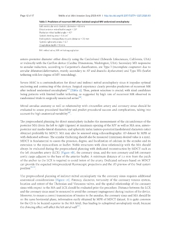
Table 2. Predictors of recurrent MR after isolated surgical MV undersized annuloplasty
Left ventricular end-diastolic diameter > 65 mm
Distal anterior mitral leaflet angle > 25°
Posterior mitral leaflet angle > 45°
Systolic tenting area > 2.5 cm 2
End-systolic interpapillary muscle distance > 20 mm
Systolic sphericity index > 0.7
Coaptation depth > 10 mm
MV: mitral valve; MR: mitral regurgitation
antero-posterior diameter either directly using the Cardioband (Edwards Lifesciences, California, USA)
or indirectly with the Carillon device (Cardiac Dimensions, Washington, USA). Secondary MR responsive
to annular reduction, according to Carpentier’s classification, are Type I (incomplete coaptation due to
annular dilatation/deformation, mainly secondary to AF and diastolic dysfunction) and Type IIIb (leaflet
tethering with low degree of MV remodeling).
Severe MAC is a contraindication for direct and indirect mitral annuloplasty since it impedes optimal
anchoring and contracting of the devices. Surgical experience clearly provides predictors of recurrent MR
after isolated undersized annuloplasty [Table 2]. Thus, patient selection is crucial, with ideal candidates
[43]
being patients with limited leaflet tethering, as suggested by high rate of recurrent MR observed in
randomized trials in surgically unselected series .
[44]
Mitral annulus anatomy as well as relationship with circumflex artery and coronary sinus should be
evaluated to assess procedural feasibility and predict procedural success and complications, taking into
[45]
account the high anatomical variability .
The preprocedural planning for direct annuloplasty includes the measurement of the circumference of the
posterior MA (from the left to right trigones) at maximum opening of the MV as well as MA area, antero-
posterior and medio-lateral diameters, and sphericity index (antero-posterior/mediolateral diameters ratio)
obtained preferably by MDCT. MA may also be assessed using echocardiographic 3D dataset by MPR or
with dedicated software. The annular thickening should also be measured (minimum desired value is 4 mm).
MDCT is fundamental to assess the presence, degree, and localization of calcium in the annulus and its
extension to the myocardium or leaflet. Noble structures with close relationship with the MA should
always be evaluated during the preprocedural planning with dedicated reconstruction by MDCT such as
the left circumflex artery (LCX) [Figure 5B], the coronary sinus, and the non-coronary and left coronary
aortic cusps adjacent to the base of the anterior leaflet. A minimum distance of 2.5 mm from the patch
of the anchor to the LCX is required to avoid lesion of the artery. Dedicated software based on MDCT
can provide the expected intraprocedural fluoroscopic projections and the 3D preview of the final system
position [32,46] .
The preprocedural planning of indirect mitral annuloplasty via the coronary sinus requires additional
anatomical considerations [Figure 10]. Patency, diameter, tortuosity of the coronary venous system,
location and extent of the Thebesian and Vieussens valves, and the spatial relationship of the coronary
sinus with respect to the MA and LCX should be evaluated prior the procedure. Distance between the LCX
and the coronary sinus must be measured to avoid the coronary impingement during traction of the device.
Moreover, to ensure a correct transmission of tension to the annulus, the coronary sinus and MA should lie
on the same horizontal plane, information easily obtained by MPR of MDCT dataset. It is quite common
for the CS to be located superior to the MA level, thus leading to suboptimal annuloplasty result, because
the chincing effect will affect the left atrial wall .
[47]