
Page 849 - Read Online
P. 849
Page 6 of 17 Melillo et al. Mini-invasive Surg 2020;4:81 I http://dx.doi.org/10.20517/2574-1225.2020.83
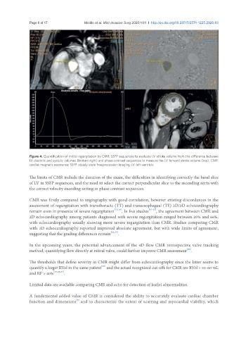
Figure 4. Quantification of mitral regurgitation by CMR. SSFP sequences to evaluate LV stroke volume from the difference between
LV diastolic and systolic volumes (bottom right) and phase contrast sequences to measure the LV forward stroke volume (top). CMR:
cardiac magnetic resonance; SSFP: steady-state free precession imaging; LV: left ventricle
The limits of CMR include the duration of the exam, the difficulties in identifying correctly the basal slice
of LV in SSFP sequences, and the need to select the correct perpendicular slice to the ascending aorta with
the correct velocity encoding setting in phase contrast sequences.
CMR was firstly compared to angiography with good correlation, however existing discordances in the
assessment of regurgitation with transthoracic (TT) and transesophageal (TE) 2D/3D echocardiography
remain even in presence of severe regurgitation [15,16] . In five studies [17-21] , the agreement between CMR and
2D echocardiography among patients diagnosed with severe regurgitation ranged between 20% and 66%,
with echocardiography usually showing more severe regurgitation than CMR. Studies comparing CMR
with 3D echocardiography reported improved absolute agreement, but with wide limits of agreement,
suggesting that the grading differences remain [22,23] .
In the upcoming years, the potential advancement of the 4D-flow CMR retrospective valve tracking
[24]
method, quantifying flow directly at mitral valve, could further improve CMR assessment .
The thresholds that define severity in CMR might differ from echocardiography since the latter seems to
[25]
quantify a larger RVol in the same patient and the actual recognized cut-offs for CMR are RVol > 55-60 mL
and RF > 40% [17,26,27] .
Limited data are available comparing CMR and echo for detection of leaflet abnormalities.
A fundamental added value of CMR is considered the ability to accurately evaluate cardiac chamber
function and dimensions and to characterize the extent of scarring and myocardial viability, which
[9]