
Page 846 - Read Online
P. 846
Melillo et al. Mini-invasive Surg 2020;4:81 I http://dx.doi.org/10.20517/2574-1225.2020.83 Page 3 of 17
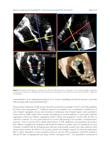
Figure 1. MPR analysis of 3D dataset to measure mitral valve area. The mitral valve is fully open. The tips of mitral leaflets are identified
in the blue and red planes to delineate the orifice, allowing the measurement of the mitral valve area on the axial. MPR : multiplanar
reconstruction
demonstrated to be an independent predictor of LV reverse remodeling and clinical outcomes in patients
[7]
with secondary MR treated with MitraClip .
Preprocedural evaluation of MR severity should be performed according to EACVI and ASE guidelines
[8,9]
for native valve regurgitation . Different methods are available such as qualitative (visualization of
color-Doppler regurgitant jet area and evaluation of continuous wave Doppler signal), semi-quantitative
(vena contracta width, mitral inflow velocity, and pulmonary vein flow pattern), or quantitative (effective
regurgitant orifice area (EROA), regurgitant volume (RVol), and regurgitant fraction (RF) by PISA or
volumetric method). As a true gold standard for accurate MR grading is not available, a multiparametric
approach is the cornerstone for a reliable quantification of MR. Qualitative and quantitative parameters
have different strengths and weaknesses that should be balanced to reduce their own limitations: jet area
is linearly correlated with the driving pressure and may underestimate eccentric jets; vena contracta width
measurement assumes the EROA to be circular; pulsed wave Doppler analysis of mitral and pulmonary
flow is highly dependent on intra-chambers pressures and left atrial compliance; and PISA method is
[9]
affected by time of regurgitation and shape of flow convergence area . The quantitative assessment of