
Page 848 - Read Online
P. 848
Melillo et al. Mini-invasive Surg 2020;4:81 I http://dx.doi.org/10.20517/2574-1225.2020.83 Page 5 of 17
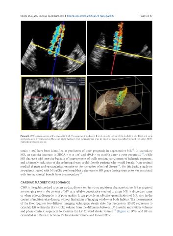
Figure 3. MPR reconstruction of the regurgitant jet. The narrowest portion of the jet close to the tip of the leaflets is identified and vena
contracta area is measured on the axial plane (yellow). The measurement may be done for each regurgitant jet and the value. MPR:
multiplanar reconstruction
[1]
strain < 2%) have been identified as predictors of poor prognosis in degenerative MR . In secondary
2
[12]
MR, an exercise increase in EROA > 0.13 cm and sPAP > 60 mmHg carry a poor prognosis , while
MR decrease with exercise because of improvement of walls motion, recruitment of ischemic segments,
and ultimately reduction of the tethering forces could identify patients who would benefit from optimal
[13]
medical therapy and revascularization prior to the correction of mitral disease . On this basis, a study on
39 patients treated with MitraClip confirmed that a decrease in MR grade during stress echo was associated
[14]
with limited clinical benefit from the procedure .
CARDIAC MAGNETIC RESONANCE
CMR is the gold standard to assess cardiac dimension, function, and tissue characterization. It has acquired
an emerging role in the context of MV as a reliable quantitative method to assess MR in discordant cases
or when echocardiography is of poor quality. It can provide an effective quantification of MR, also in the
contest of multivalvular disease, without limitations of imaging window or body habitus. The measurement
of the Rvol requires two different imaging techniques: steady state free precession (SSFP) sequences to
calculate left ventricular (LV) stroke volume from the difference between LV diastolic and systolic volumes
and phase contrast sequences to measure the LV forward stroke volume [Figure 4]. RVol and RF are
[15]
calculated as difference between LV total stroke volume and forward flow.