
Page 188 - Read Online
P. 188
Jairath et al. Percutaneous nephrostomy
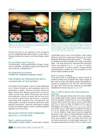
Figure 1: The patient in prone position with roller pack underneath Figure 2: Surface marking (concept of Quadrangle of safety): with
upper abdomen and chest, abdominal contents falls forward so as the patient in prone position, Quadrangle of safety is formed by
to give proper access to the kidneys posterior axillary line as lateral limit, upper margin of iliac crest as
lower limit, lateral margin of paraspinous muscle as medial limit, the
11th and 12th rib border as upper limit
Urinary diversion in an attempt to heal conditions
such as malignant/inflammatory fistula, urinary leak or
fistulas resulting from trauma, and hemorrhagic cystitis underneath pelvic bone and another under upper
etc. [2,3] . abdomen and chest region (as shown) so as to give
adequate stretching around flank region [4,5] . The side to
be operated should be brought at the edge of operating
For providing route of access
Chemotherapy, antifungal/antibiotic therapy, benign table. The area should be cleansed with povidone
stricture dilatation, antegrade ureteral stent placement, iodine and draped [Figure 1]. In case of relative
contraindication to prone position (compromised
stone retrieval, endopyelotomy [2,3] .
cardiorespiratory system etc.), this procedure can be
done in supine position as well.
For diagnostic procedures
[3]
Whitaker test, antegrade pyelography, biopsy .
Step 2: surface marking
If we place PCN in quadrangle of safety formed by
PRE-OPERATIVE PREPARATION AND A posterior axillary line as lateral limit, upper margin of
COUNSELING OF THE PATIENT iliac crest as lower limit, lateral margin of paraspinous
muscle as medial limit, the 11th and 12th rib border
Commonly, this procedure is done in local anesthesia as upper limit, there are less chances of associated
(LA). Patient should be well explained about the intrabdominal visceral injuries [Figure 2] [4,5] .
procedure in detail. Informed consent should be
taken beforehand. Bleeding parameters should be Step 3: USG to decide site of percutaneous
within normal limits. Attain intravenous (IV) access puncture
and antibiotics should be given half an hour prior USG of the diseased kidney should be done starting
to procedure particularly particularly in patients from medial aspect (Para spinal), advancing laterally
presenting with urosepsis. For uncooperative but until the posterior axillary line so as to see posterior
willing patient, procedure should be performed under calyces first followed by lateral calyces thereafter
general anesthesia. Relevant radiological images and thus to have an idea of degree of HN, type of
should be reviewed again in order to decide anoptimal pathology in the renal unit [Figure 3A and B]. We in
approach for renal access. our institute use 3.5 MHz convex transducer focused at
5-9 cm for adults and 5 MHz transducer focused at
DETAILED STEP-BY-STEP NUMBERED 5-7 cm for children. Exact site of puncture depends
primarily on the cause of hydronephrosis (HDN) and
MEDICAL ILLUSTRATION anatomic landmarks. For simple urinary drainage a
lower pole posterior calyx is usually best which can
Step 1: patient positioning be easily accessed via subcoastal approach. For
With patient in prone position, a roller pack is placed accessing pelvicureteric junction (PUJ) or upper ureter,
Mini-invasive Surgery ¦ Volume 1 ¦ December 28, 2017 181