Page 176 - Read Online
P. 176
De Aquino et al.                                                                                                                                              Esophageal mucosectomy in advanced achalasia

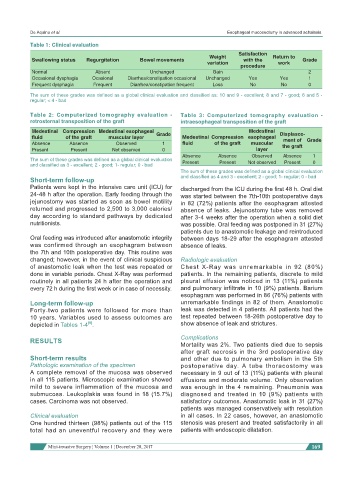
           Table 1: Clinical evaluation
                                                                                 Satisfaction
                                                                        Weight               Return to
            Swallowing status  Regurgitation    Bowel movements                    with the            Grade
                                                                       variation               work
                                                                                  procedure
            Normal               Absent             Unchanged            Gain                            2
            Occasional dysphagia  Ocasional  Diarrhea/constipation occasional  Unchanged  Yes  Yes       1
            Frequent dysphagia  Frequent     Diarrhea/constipation frequent  Loss    No         No       0
           The sum of these grades was defined as a global clinical evaluation and classified as: 10 and 9 - excellent; 8 and 7 - good; 6 and 5 -
           regular; < 4 - bad


           Table 2:  Computerized tomography evaluation -     Table 3:  Computerized tomography evaluation -
           retrosternal transposition of the graft            intraesophageal transposition of the graft
            Medestinal  Compression  Medestinal esophageal   Grade                   Medestinal   Displasce-
            fluid      of the graft   muscular layer           Medestinal  Compression  esophageal   ment of  Grade
            Absence     Absence         Observed        1      fluid     of the graft  muscular   the graft
            Present      Present       Not observed     0                              layer
                                                               Absence     Absence    Observed   Absence   1
           The sum of these grades was defined as a global clinical evaluation   Present  Present  Not observed  Present  0
           and classified as 3 - excellent; 2 - good; 1- regular; 0 - bad
                                                              The sum of these grades was defined as a global clinical evaluation
           Short-term follow-up                               and classified as 4 and 3 - excellent; 2 - good; 1- regular; 0 - bad
           Patients were kept in the intensive care unit (ICU) for   discharged from the ICU during the first 48 h. Oral diet
           24-48 h after the operation. Early feeding through the   was started between the 7th-10th postoperative days
           jejunostomy was started as soon as bowel motility   in 82 (72%) patients after the esophagram attested
           returned and progressed to 2,500 to 3,000 calories/  absence of leaks. Jejunostomy tube was removed
           day according to standard pathways by dedicated    after 3-4 weeks after the operation when a solid diet
           nutritionists.                                     was possible. Oral feeding was postponed in 31 (27%)
                                                              patients due to anastomotic leakage and reintroduced
           Oral feeding was introduced after anastomotic integrity   between days 18-29 after the esophagram attested
           was confirmed through an esophagram between        absence of leaks.
           the 7th and 10th postoperative day. This routine was
           changed; however, in the event of clinical suspicious   Radiologic evaluation
           of anastomotic leak when the test was repeated or   Chest X-Ray was unremarkable in 92 (80%)
           done in variable periods. Chest X-Ray was performed   patients. In the remaining patients, discrete to mild
           routinely in all patients 24 h after the operation and   pleural effusion was noticed in 13 (11%) patients
           every 72 h during the first week or in case of necessity.  and pulmonary infiltrate in 10 (9%) patients. Barium
                                                              esophagram was performed in 86 (76%) patients with
           Long-term follow-up                                unremarkable findings in 82 of them. Anastomotic
           Forty-two patients were followed for more than     leak was detected in 4 patients. All patients had the
           10 years. Variables used to assess outcomes are    test repeated between 18-26th postoperative day to
                               [9]
           depicted in Tables 1-4 .                           show absence of leak and strictures.

           RESULTS                                            Complications
                                                              Mortality was 2%. Two patients died due to sepsis
                                                              after graft necrosis in the 3rd postoperative day
           Short-term results                                 and other due to pulmonary embolism in the 5th
           Pathologic examination of the specimen             postoperative day. A tube thoracostomy was
           A complete removal of the mucosa was observed      necessary in 9 out of 13 (11%) patients with pleural
           in all 115 patients. Microscopic examination showed   effusions and moderate volume. Only observation
           mild to severe inflammation of the mucosa and      was enough in the 4 remaining. Pneumonia was
           submucosa. Leukoplakia was found in 18 (15.7%)     diagnosed  and  treated  in  10  (9%)  patients  with
           cases. Carcinoma was not observed.                 satisfactory outcomes. Anastomotic leak in 31 (27%)
                                                              patients was managed conservatively with resolution
           Clinical evaluation                                in all cases. In 22 cases, however, an anastomotic
           One hundred thirteen (98%) patients out of the 115   stenosis was present and treated satisfactorily in all
           total had an uneventful recovery and they were     patients with endoscopic dilatation.

                          Mini-invasive Surgery ¦ Volume 1 ¦ December 28, 2017                            169

