
Page 94 - Read Online
P. 94
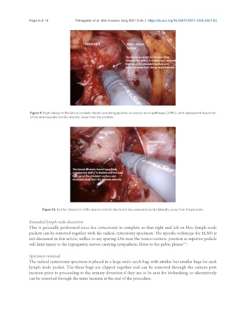
Page 8 of 18          Thinagaran et al. Mini-invasive Surg 2021;5:46  https://dx.doi.org/10.20517/2574-1225.2021.53


                Figure 9. High release of the lateral prostatic fascia containing putative accessory nerve pathways (ANPs), with subsequent dissection
                of the neurovascular bundle laterally, away from the prostate.


                      Figure 10. Further dissection of the lateral prostatic fascia and neurovascular bundle laterally, away from the prostate.

               Extended lymph node dissection
               This is generally performed once the cystectomy is complete so that right and left en bloc lymph node
               packets can be removed together with the radical cystectomy specimen. The specific technique for ELND is
               not discussed in this article; suffice to say sparing LNs near the vesico-ureteric junction at superior pedicle
               will limit injury to the hypogastric nerves carrying sympathetic fibres to the pelvic plexus .
                                                                                          [17]
               Specimen removal
               The radical cystectomy specimen is placed in a large endo catch bag, with similar but smaller bags for each
               lymph node packet. The three bags are clipped together and can be removed through the camera port
               incision prior to proceeding to the urinary diversion if they are to be sent for biobanking, or alternatively
               can be removed through the same incision at the end of the procedure.