
Page 90 - Read Online
P. 90
Page 4 of 18 Thinagaran et al. Mini-invasive Surg 2021;5:46 https://dx.doi.org/10.20517/2574-1225.2021.53
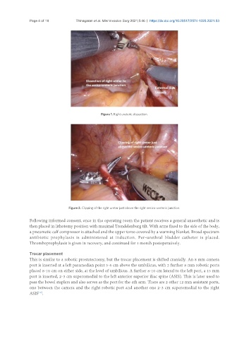
Figure 1. Right ureteric dissection.
Figure 2. Clipping of the right ureter just above the right vesico-ureteric junction.
Following informed consent, once in the operating room the patient receives a general anaesthetic and is
then placed in lithotomy position with maximal Trendelenburg tilt. With arms fixed to the side of the body,
a pneumatic calf compressor is attached and the upper torso covered by a warming blanket. Broad spectrum
antibiotic prophylaxis is administered at induction. Per-urethral bladder catheter is placed.
Thromboprophylaxis is given in recovery, and continued for 1 month postoperatively.
Trocar placement
This is similar to a robotic prostatectomy, but the trocar placement is shifted cranially. An 8 mm camera
port is inserted at a left paramedian point 5-6 cm above the umbilicus, with 2 further 8 mm robotic ports
placed 8-10 cm on either side, at the level of umbilicus. A further 8-10 cm lateral to the left port, a 15 mm
port is inserted, 2-3 cm superomedial to the left anterior superior iliac spine (ASIS). This is later used to
pass the bowel staplers and also serves as the port for the 4th arm. There are 2 other 12 mm assistant ports,
one between the camera and the right robotic port and another one 2-3 cm superomedial to the right
ASIS .
[12]