
Page 93 - Read Online
P. 93
Thinagaran et al. Mini-invasive Surg 2021;5:46 https://dx.doi.org/10.20517/2574-1225.2021.53 Page 7 of 18
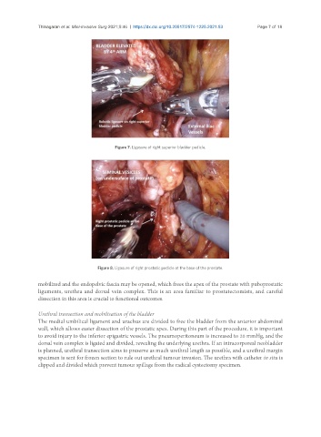
Figure 7. Ligasure of right superior bladder pedicle.
Figure 8. Ligasure of right prostatic pedicle at the base of the prostate.
mobilized and the endopelvic fascia may be opened, which frees the apex of the prostate with puboprostatic
ligaments, urethra and dorsal vein complex. This is an area familiar to prostatectomists, and careful
dissection in this area is crucial to functional outcomes.
Urethral transection and mobilisation of the bladder
The medial umbilical ligament and urachus are divided to free the bladder from the anterior abdominal
wall, which allows easier dissection of the prostatic apex. During this part of the procedure, it is important
to avoid injury to the inferior epigastric vessels. The pneumoperitoneum is increased to 20 mmHg, and the
dorsal vein complex is ligated and divided, revealing the underlying urethra. If an intracorporeal neobladder
is planned, urethral transection aims to preserve as much urethral length as possible, and a urethral margin
specimen is sent for frozen section to rule out urethral tumour invasion. The urethra with catheter in situ is
clipped and divided which prevent tumour spillage from the radical cystectomy specimen.