
Page 55 - Read Online
P. 55
Jacoby et al. Mini-invasive Surg 2022;6:58 https://dx.doi.org/10.20517/2574-1225.2022.58 Page 7 of 11
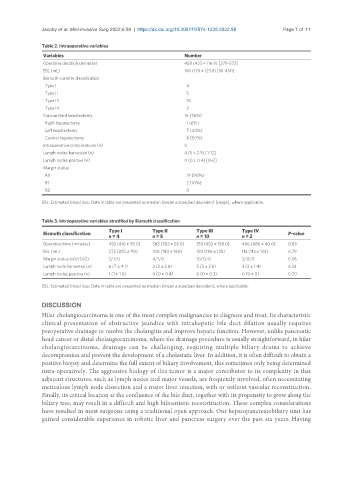
Table 2. Intraoperative variables
Variables Number
Operative duration (minutes) 458 (433 ± 116.9) [279-573]
EBL (mL) 150 (175 ± 123.8) [10-450]
Bismuth-corlette classification
Type I 4
Type II 5
Type III 10
Type IV 2
Concomitant hepatectomy 16 (76%)
Right hepatectomy 1 (6%)
Left hepatectomy 7 (44%)
Central hepatectomy 8 (50%)
Intraoperative complications (n) 0
Lymph nodes harvested (n) 4 (5 ± 2.9) [1-12]
Lymph nodes positive (n) 0 (0 ± 0.4) [0-2]
Margin status
R0 19 (90%)
R1 2 (10%)
R2 0
EBL: Estimated blood loss; Data in table are presented as median (mean ± standard deviation) [range], where applicable.
Table 3. Intraoperative variables stratified by Bismuth classification
Type I Type II Type III Type IV
Bismuth classification n = 4 n = 5 n = 10 n = 2 P-value
Operative time (minutes) 453 (416 ± 93.0) 383 (383 ± 55.0) 518 (453 ± 158.0) 486 (486 ± 40.0) 0.69
EBL (mL) 275 (225 ± 119) 100 (180 ± 160) 150 (166 ± 125) 113 (113 ± 124) 0.79
Margin status (r0/r1/r2) 3/1/0 4/1/0 10/0/0 2/0/0 0.36
Lymph node harvested (n) 6 (7 ± 4.1) 3 (3 ± 2.6) 5 (5 ± 2.6) 3 (3 ± 1.4) 0.33
Lymph nodes positive (n) 1 (1 ± 1.0) 0 (0 ± 0.4) 0 (0 ± 0.3) 0 (0 ± 0) 0.20
EBL: Estimated blood loss; Data in table are presented as median (mean ± standard deviation), where applicable.
DISCUSSION
Hilar cholangiocarcinoma is one of the most complex malignancies to diagnose and treat. Its characteristic
clinical presentation of obstructive jaundice with intrahepatic bile duct dilation usually requires
preoperative drainage to resolve the cholangitis and improve hepatic function. However, unlike pancreatic
head cancer or distal cholangiocarcinoma, where the drainage procedure is usually straightforward, in hilar
cholangiocarcinoma, drainage can be challenging, requiring multiple biliary drains to achieve
decompression and prevent the development of a cholestatic liver. In addition, it is often difficult to obtain a
positive biopsy and determine the full extent of biliary involvement, this sometimes only being determined
intra-operatively. The aggressive biology of this tumor is a major contributor to its complexity in that
adjacent structures, such as lymph nodes and major vessels, are frequently involved, often necessitating
meticulous lymph node dissection and a major liver resection, with or without vascular reconstruction.
Finally, its critical location at the confluence of the bile duct, together with its propensity to grow along the
biliary tree, may result in a difficult and high bilioenteric reconstruction. These complex considerations
have resulted in most surgeons using a traditional open approach. Our hepatopancreatobiliary unit has
gained considerable experience in robotic liver and pancreas surgery over the past six years. Having