
Page 51 - Read Online
P. 51
Jacoby et al. Mini-invasive Surg 2022;6:58 https://dx.doi.org/10.20517/2574-1225.2022.58 Page 3 of 11
according to the Clavien-Dindo classification, length of stay (LOS), 30-day readmission, in-hospital
mortality, 90-day mortality, follow-up time, and current oncological status.
The preoperative workup included a triple phase 1-mm cut computed tomography (CT) scan of the
abdomen and pelvis, chest CT, and abdominal magnetic resonance imaging (MRI) with magnetic resonance
cholangiopancreatography (MRCP). This high-quality imaging is mainly performed to evaluate the blood
vessels (portal vein and hepatic artery) along the porta hepatis and to assess any aberrant vasculature and/or
vessel involvement. MRCP is performed to map the biliary tree, assess the extent of intrahepatic sectoral
biliary obstruction, and plan reconstruction of the biliary tree after completion of resection. Future liver
remnant volume was calculated on the basis of imaging findings in patients who required extended major
hepatic resection. Patients with cholangitis, hyperbilirubinemia > 3 mg/dL, or who required a major
hepatectomy with borderline future liver remnant volume underwent preoperative biliary drainage. In most
cases, drainage was achieved by endoscopic retrograde cholangiopancreatography (ERCP) with brushing
and stent placement. Many of the patients had already undergone this procedure prior to referral to our
center. When available and necessary, cholangioscopy with biopsy was added. Percutaneous transhepatic
cholangiography (PTC) was performed when bilirubin concentrations remained high following ERCP
stenting or when ERCP was not technically feasible. In addition, every patient was clinically evaluated in
terms of performance status, medical comorbidities, and cardiac and liver function.
Operative procedure
®
All operations were performed using the da Vinci Xi robotic surgical system (Intuitive Surgical, Sunnyvale,
CA, USA). Exclusion criteria for the robotic approach comprised hepatic artery and/or main portal vein
invasion that required major vascular resection and reconstruction. Patients were placed in a supine
position with both hands abducted to less than 90° on arm boards. Following the insertion of an 8-mm port
through the umbilicus, the procedure commenced with diagnostic laparoscopy. After excluding peritoneal
spread, an 8-mm port was placed along the right midclavicular line and a 12-mm port along the left
midclavicular line, both at the level of the umbilicus. A fourth 8-mm port was placed in the left anterior
®
axillary line. Finally, an Advanced Access Gelport (Applied Medical, Rancho Santa Margarita, CA, USA)
was placed between the right midclavicular line and umbilicus, slightly inferior to the umbilicus. The
®
Gelport is used by the bedside surgeon, mainly for suctioning and insertion of sutures. The incision
(3-5 cm) needed for the Gelport is also used as the extraction site for the specimen. An AirSeal port
®
®
(ConMed, Largo, FL, USA) was inserted through the Gelport for insufflation and smoke evacuation
®
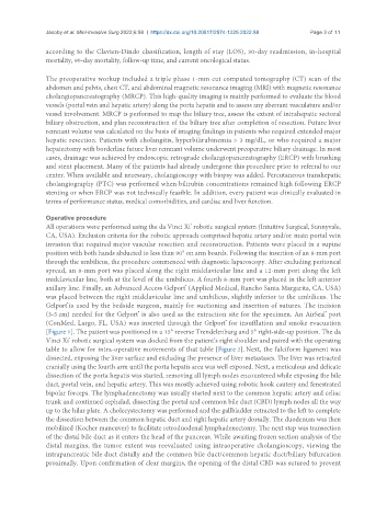
[Figure 1]. The patient was positioned in a 15° reverse Trendelenburg and 5° right-side-up position. The da
Vinci Xi robotic surgical system was docked from the patient’s right shoulder and paired with the operating
®
table to allow for intra‐operative movements of that table [Figure 2]. Next, the falciform ligament was
dissected, exposing the liver surface and excluding the presence of liver metastases. The liver was retracted
cranially using the fourth arm until the porta hepatis area was well exposed. Next, a meticulous and delicate
dissection of the porta hepatis was started, removing all lymph nodes encountered while exposing the bile
duct, portal vein, and hepatic artery. This was mostly achieved using robotic hook cautery and fenestrated
bipolar forceps. The lymphadenectomy was usually started next to the common hepatic artery and celiac
trunk and continued cephalad, dissecting the portal and common bile duct (CBD) lymph nodes all the way
up to the hilar plate. A cholecystectomy was performed and the gallbladder retracted to the left to complete
the dissection between the common hepatic duct and right hepatic artery dorsally. The duodenum was then
mobilized (Kocher maneuver) to facilitate retroduodenal lymphadenectomy. The next step was transection
of the distal bile duct as it enters the head of the pancreas. While awaiting frozen section analysis of the
distal margins, the tumor extent was reevaluated using intraoperative cholangioscopy, viewing the
intrapancreatic bile duct distally and the common bile duct/common hepatic duct/biliary bifurcation
proximally. Upon confirmation of clear margins, the opening of the distal CBD was sutured to prevent