
Page 52 - Read Online
P. 52
Page 4 of 11 Jacoby et al. Mini-invasive Surg 2022;6:58 https://dx.doi.org/10.20517/2574-1225.2022.58
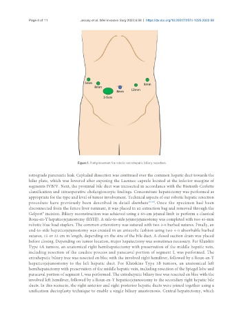
Figure 1. Port placement for robotic extrahepatic biliary resection.
retrograde pancreatic leak. Cephalad dissection was continued over the common hepatic duct towards the
hilar plate, which was lowered after exposing the Laennec capsule located at the inferior margins of
segments IVB/V. Next, the proximal bile duct was transected in accordance with the Bismuth-Corlette
classification and intraoperative cholangioscopic findings. Concomitant hepatectomy was performed as
appropriate for the type and level of tumor involvement. Technical aspects of our robotic hepatic resection
procedure have previously been described in detail elsewhere [15,16] . Once the specimen had been
disconnected from the future liver remnant, it was placed in an extraction bag and removed through the
Gelport® incision. Biliary reconstruction was achieved using a 60-cm jejunal limb to perform a classical
Roux-en-Y hepaticojejunostomy (RYHJ). A side-to-side jejunojejunostomy was completed with two 45-mm
robotic blue load staplers. The common enterotomy was sutured with two 3‐0 barbed sutures. Finally, an
end-to-side hepaticojejunostomy was created in an antecolic fashion using two 4-0 absorbable barbed
sutures, 15 or 22 cm in length, depending on the size of the bile duct. A closed suction drain was placed
before closing. Depending on tumor location, major hepatectomy was sometimes necessary. For Klatskin
Type 3A tumors, an anatomical right hemihepatectomy with preservation of the middle hepatic vein,
including resection of the caudate process and paracaval portion of segment I, was performed. The
extrahepatic biliary tree was resected en bloc with the involved right hemiliver, followed by a Roux-en-Y
hepaticojejunostomy to the left hepatic duct. For Klatskins Type 3B tumors, an anatomical left
hemihepatectomy with preservation of the middle hepatic vein, including resection of the Spiegel lobe and
paracaval portion of segment I, was performed. The extrahepatic biliary tree was resected en bloc with the
involved left hemiliver, followed by a Roux-en-Y hepaticojejunostomy to the secondary right hepatic bile
ducts. In this scenario, the right anterior and right posterior hepatic ducts were joined together using a
unification ductoplasty technique to enable a single biliary anastomosis. Central hepatectomy, which