
Page 54 - Read Online
P. 54
Page 6 of 11 Jacoby et al. Mini-invasive Surg 2022;6:58 https://dx.doi.org/10.20517/2574-1225.2022.58
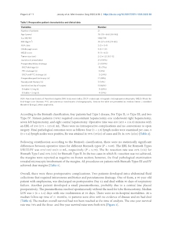
Table 1. Preoperative patient characteristics and clinical data
Variables Number
Number of patients 21
Age (years) 72 (72 ± 8.6) [55-90]
Sex (M/W) 14M/7W
2
BMI (kg/m ) 25 (27 ± 5.9) [19-40]
ASA class 3 (3 ± 0.4)
Childs-pugh score 5 (6 ± 1.0)
MELD score 9 (11 ± 6.0)
Tumor size (cm) 2 (2 ± 1.2) [0.7-3]
Jaundice at presentation 21 (100%)
Preoperative biliary drainage 21 (100%)
ERCP drainage (n) 15 (71%)
PTC drainage (n) 1 (5%)
ERCP and PTC drainage (n) 5 (24%)
Preoperative positive biopsy (n) 17 (81%)
Neoadjuvant therapy (n) 3 (14%)
Jaundiced on day of surgery 13 (62%)
Bilirubin 1-3 mg/dL 9 (69%)
Bilirubin > 3 mg/dL 4 (31%)
ASA: American Society of Anesthesiologists; BMI: body mass index; ERCP: endoscopic retrograde cholangiopancreatography; MELD: Model for
End-Stage Liver Disease; PTC: percutaneous transhepatic cholangiography. Data in the table are presented as median (mean ± standard
deviation) [range], where applicable.
According to the Bismuth classification, four patients had Type I disease, five Type II, 10 Type III, and two
Type IV. Sixteen patients (76%) required concomitant hepatectomy; one underwent right hepatectomy,
seven left hepatectomy, and eight central hepatectomy. Operative time was 458 (433 ± 116.9) minutes with
an EBL of 150 (175 ± 123.8) mL. There were no intraoperative complications and no conversions to open
surgery. Final pathological outcomes were as follows: four (5 ± 2.9) lymph nodes were examined per case, 0
(0 ± 0.4) lymph nodes were positive, R0 was attained in 90% (19/21) of cases and R1 in 10% (2/21) [Table 2].
Following stratification according to the Bismuth classification, there were no statistically significant
differences between operative times for different Bismuth types (P = 0.69). The EBL for Bismuth Types
I/II/III/IV was 275/100/150/113 mL, respectively (P = 0.79). The R1 resection rate was 25% (1/4) for
Bismuth Type I and 20% (1/5) for Bismuth Type II. In the two cases in which R1 resection was not achieved,
the margins were reported as negative on frozen section; however, the final pathological examination
revealed microscopic involvement of the margins. All procedures on patients with Bismuth Types III and IV
achieved clear margins [Table 3].
Overall, there were three postoperative complications. Two patients developed intra-abdominal fluid
collections that required intravenous antibiotics and percutaneous drainage. One of them, a 90-year-old
patient with emphysema, was discharged on postoperative Day 12 and died within 90 days of respiratory
failure. Another patient developed a small pneumothorax, probably due to a central line placed
preoperatively. The pneumothorax resolved spontaneously without the need for tube thoracostomy. Median
LOS was 5 (6 ± 3.4) days with one readmission at 30 days. There were no in-hospital mortalities. At a
median follow-up time of 21 months, 15 patients were alive with no evidence of disease and six had died
[Table 4]. The median overall survival had not been reached at the time of analysis. The one-year survival
rate was 78% and the three- and five-year survival rates were both 60% [Figure 3].