
Page 53 - Read Online
P. 53
Jacoby et al. Mini-invasive Surg 2022;6:58 https://dx.doi.org/10.20517/2574-1225.2022.58 Page 5 of 11
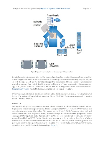
Figure 2. Operation room setup for robotic extrahepatic biliary resection.
included resection of segments 4B/5 and the paracaval portion of the caudate lobe, was only performed for
Klatskin Type 2 tumors with limited involvement of the biliary bifurcation after securing negative margins
on both the right and left hepatic ducts by intraoperative examination of frozen sections. This situation is
much less common than typical Klatskin tumors, and in most of them, preoperative biliary endoscopy
®
SpyGlass (Boston Scientific Corporation, Natick, MA, USA) suggested limited tumor involvement.
Supplementary video 1 attached to this manuscript depicts our surgical procedure.
Data were documented on an Excel (Microsoft) spreadsheet and analyses were carried out using GraphPad
Prism 8TM software (GraphPad Software, San Diego, CA, USA). The data are presented as median
(mean ± standard deviation).
RESULTS
During the study period, 21 patients underwent robotic extrahepatic biliary resection, with or without
hepatectomy, for hilar cholangiocarcinoma. The median age was 72 (71 ± 8.3) years; 14 (67%) were men and
2
seven (33%) were women. The median BMI was 25 (27 ± 5.9) kg/m , Child-Pugh score 5 (6 ± 1.0), and
MELD score 9 (11 ± 6.0). All patients initially presented with jaundice and underwent preoperative biliary
drainage; 15 (71%) patients had a stent placed by ERCP, one (5%) was stented by PTC, and five (24%)
required both ERCP and PTC. Positive biopsies were obtained in 17 (81%) patients, three (24%) of whom
were referred for neoadjuvant treatment before resection. On the day of operation, 13 (62%) patients had
persistent, mostly mild, hyperbilirubinemia (1-3 mg/dL). Four patients had persistent hyperbilirubinemia
with bilirubin > 3 mg/dL despite all drainage efforts [Table 1].