
Page 16 - Read Online
P. 16
Saleh et al. J Transl Genet Genom 2021;5:250-64 https://dx.doi.org/10.20517/jtgg.2021.23 Page 260
Table 3. Correlation of ABCA4 genotype with lesions detected only by W-OCT
Group Allel 1/Allel 2 No. of patients without lesions (mean age ± SD) No. of patients with lesions (mean age ± SD)
1 Pathogenic/+ 2 (27.95 ± 26.09 year) 2 (48.6 ± 28.43 year)
2 Pathogenic/mild 13 (32.15 ± 5.76 year) 7 (48.37 ± 9.79 year)
3 Pathogenic/pathogenic - 28 (34.10 ± 15.47 year)
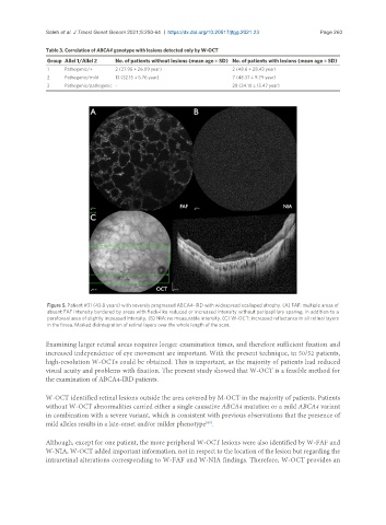
Figure 5. Patient #31 (43.8 years) with severely progressed ABCA4-IRD with widespread scalloped atrophy. (A) FAF: multiple areas of
absent FAF intensity bordered by areas with fleck-like reduced or increased intensity without peripapillary sparing, in addition to a
parafoveal area of slightly increased intensity. (B) NIA: no measurable intensity. (C) W-OCT: increased reflectance in all retinal layers
in the fovea. Marked disintegration of retinal layers over the whole length of the scan.
Examining larger retinal areas requires longer examination times, and therefore sufficient fixation and
increased independence of eye movement are important. With the present technique, in 50/52 patients,
high-resolution W-OCTs could be obtained. This is important, as the majority of patients had reduced
visual acuity and problems with fixation. The present study showed that W-OCT is a feasible method for
the examination of ABCA4-IRD patients.
W-OCT identified retinal lesions outside the area covered by M-OCT in the majority of patients. Patients
without W-OCT abnormalities carried either a single causative ABCA4 mutation or a mild ABCA4 variant
in combination with a severe variant, which is consistent with previous observations that the presence of
mild alleles results in a late-onset and/or milder phenotype .
[40]
Although, except for one patient, the more peripheral W-OCT lesions were also identified by W-FAF and
W-NIA, W-OCT added important information, not in respect to the location of the lesion but regarding the
intraretinal alterations corresponding to W-FAF and W-NIA findings. Therefore, W-OCT provides an