
Page 17 - Read Online
P. 17
Page 261 Saleh et al. J Transl Genet Genom 2021;5:250-64 https://dx.doi.org/10.20517/jtgg.2021.23
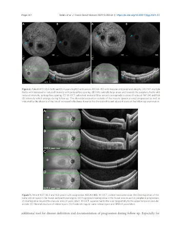
Figure 6. Patient #12 [22.6 (left) and 26.4 years (right)] with severe ABCA4-IRD with macular and peripheral atrophy. (A) FAF: multiple
flecks with increased or reduced intensity with peripapillary sparing. (B) NIA: centrally large areas and towards the periphery flecks with
reduced intensity, peripapillary sparing. (C) W-OCT: subretinal material (blue arrow) corresponds to areas of reduced FAF (A) and NIA
(B) intensity which enlarge during follow-up. The choroidal excavation outside of the macula (green arrows) progressed as well as
indicated by the absence of the line of increased reflectance towards the choroid in this and adjacent scans at the follow-up examination.
Figure 7. Patient #27 (14.3 and 18.8 years) with progressive ABCA4-IRD. W-OCT, central horizontal scan: (A) Disintegration of the
outer retinal layers in the foveal and parafoveal region. (B) Progressed disintegration in the foveal area as well as peripheral progression
of disintegration beyond the macular area (4 years later). W-OCT, superior horizontal scan tangentially to the upper temporal vascular
arcade: (C) Normal structure of retinal layers. (D) Flecks of irregular outer retinal layers and SRM (4 years later).
additional tool for disease definition and documentation of progression during follow up. Especially for