
Page 15 - Read Online
P. 15
Page 259 Saleh et al. J Transl Genet Genom 2021;5:250-64 https://dx.doi.org/10.20517/jtgg.2021.23
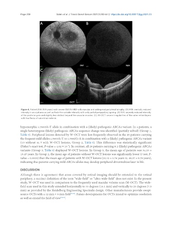
Figure 4. Patient #46 (8.8 years) with severe ABCA4-IRD with macular and widespread peripheral atrophy. (A) FAF: centrally reduced
intensity in an oval area as well as fleck-like variable intensity with only partial peripapillary sparing. (B) NIA: severely reduced intensity
at the posterior pole and slightly less distinct beyond the vascular arcades. (C) W-OCT: severe irregularities of the outer retinal layers
with few flecks of subretinal material.
hypomorphic c.5603A>T allele in combination with a (likely) pathogenic ABCA4 variant. In 4 patients, a
single heterozygous (likely) pathogenic ABCA4 sequence change was identified (partially solved) (Group 1,
Table 3). Peripheral lesions detected by W-OCT were less frequently observed in the 20 patients carrying
the frequent mild alleles c.5603A>T or c.5882G>A in combination with a (likely) pathogenic ABCA4 variant
(13 without vs. 7 with W-OCT lesions, Group 2, Table 3). This difference was statistically significant
-7
(Fisher’s exact test, P-value = 4.02 × 10 ). In contrast, all 28 patients carrying ≥ 2 (likely) pathogenic ABCA4
variants (Group 3, Table 3) displayed W-OCT lesions. In Group 3, the mean age of patients was 34.10 ±
15.47 years. In Group 2, the mean age of patients without W-OCT lesions was significantly lower (t test, P-
value = 0.0035) than the mean age of patients with W-OCT lesions (32.15 ± 5.76 years vs. 48.37 ± 9.79 years),
indicating that patients carrying mild ABCA4 alleles may develop peripheral abnormalities later in life.
DISCUSSION
Although there is agreement that areas covered by retinal imaging should be extended to the retinal
periphery, a succinct definition of the term “wide-field” or “ultra-wide-field” does not exist. In the present
study, W-OCT was used in comparison to the frequently used macular volume scan (M-OCT). The wide-
field scan used in this study extended horizontally to 55 degrees (16.1 mm) and vertically to 25 degrees (7.3
mm) as provided by the Heidelberg Engineering Spectralis design. Other manufacturers provide swept-
source OCTs with a 12 mm × 9 mm field [20,30] . Future developments for OCTs intend to optimize resolution
as well as extend the field of view [38,39] .