
Page 13 - Read Online
P. 13
Page 257 Saleh et al. J Transl Genet Genom 2021;5:250-64 https://dx.doi.org/10.20517/jtgg.2021.23
47 35.2 1 A A RI RI NoPS A R NoPS
48 33.7 0 A, FE* Normal RI Normal NPI FR Normal NPI
49 38.1 0 SRM SRM A, FRI A, FRI NoPS A, FRI FR NoPS
50 10.7 0 A, SRM SRM FRI FRI NoPS FR FR NoPS
51 60.8 0 A, SRM SRM FRI FI PS FRI FRI NoPS
52 55.4 0 SRM A*, SRM FI FI PS FRI FR PS
Optical coherence tomography (OCT) - A: areas of outer layer atrophy; FC: foveal cavitation; FE: focal choroidal excavation; PRI: preserved foveal
island; SRM: subretinal material; WA: widespread areas of outer layer atrophy. Fundus autofluorescence (FAF)/near-infrared autofluorescence
(NIA) - A: areas of absent FAF/NIA intensity; FI: flecks with increased intensity; FR: flecks with reduced intensity; FRI: flecks with increased or
reduced intensity; MR: markedly reduced intensity; ND: not done; NPI: no peripapillary involvement; NoPS: absence of peripapillary sparing; PS:
peripapillary sparing; R: generally reduced intensity; RI: ring with increased intensity; RR: ring with reduced intensity; *indicates unilateral findings.
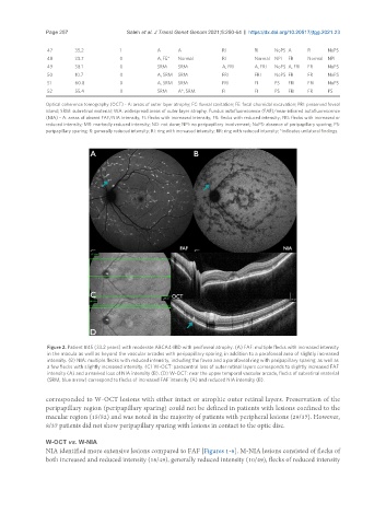
Figure 2. Patient #45 (33.2 years) with moderate ABCA4-IRD with perifoveal atrophy. (A) FAF: multiple flecks with increased intensity
in the macula as well as beyond the vascular arcades with peripapillary sparing, in addition to a parafoveal area of slightly increased
intensity. (B) NIA: multiple flecks with reduced intensity, including the fovea and a parafoveal ring with peripapillary sparing, as well as
a few flecks with slightly increased intensity. (C) W-OCT: paracentral loss of outer retinal layers corresponds to slightly increased FAF
intensity (A) and a marked loss of NIA intensity (B). (D) W-OCT: near the upper temporal vascular arcade, flecks of subretinal material
(SRM, blue arrow) correspond to flecks of increased FAF intensity (A) and reduced NIA intensity (B).
corresponded to W-OCT lesions with either intact or atrophic outer retinal layers. Preservation of the
peripapillary region (peripapillary sparing) could not be defined in patients with lesions confined to the
macular region (15/52) and was noted in the majority of patients with peripheral lesions (29/37). However,
8/37 patients did not show peripapillary sparing with lesions in contact to the optic disc.
W-OCT vs. W-NIA
NIA identified more extensive lesions compared to FAF [Figures 1-6]. M-NIA lesions consisted of flecks of
both increased and reduced intensity (16/49), generally reduced intensity (10/49), flecks of reduced intensity