
Page 14 - Read Online
P. 14
Saleh et al. J Transl Genet Genom 2021;5:250-64 https://dx.doi.org/10.20517/jtgg.2021.23 Page 258
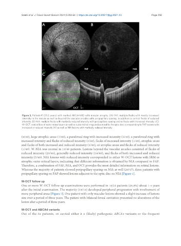
Figure 3. Patient #1 (29.5 years) with marked ABCA4-IRD with macular atrophy. (A) FAF: multiple flecks with mostly increased
intensity in the macula as well as beyond the vascular arcades with peripapillary sparing, in addition to central flecks of reduced
intensity. (B) NIA: multiple flecks with markedly reduced intensity with peripapillary sparing and no flecks with increased intensity. (C)
W-OCT: central loss of outer retinal layers as well as outer retinal irregularities nasal to the optic disc corresponding to FAF lesions with
increased or reduced intensity (A) as well as NIA lesions with markedly reduced intensity.
(8/49), large atrophic areas (7/49), a parafoveal ring with increased intensity (3/49), a parafoveal ring with
increased intensity and flecks of reduced intensity (2/49), flecks of increased intensity (1/49), atrophic areas
and flecks of both increased and reduced intensity (1/49), or atrophic areas and flecks of reduced intensity
(1/49). W-NIA was normal in 10/49 patients. Lesions beyond the vascular arcades consisted of flecks of
reduced intensity (20/49), generally reduced intensity (14/49), and flecks of both increased and reduced
intensity (5/49). NIA lesions with reduced intensity corresponded to either W-OCT lesions with SRM or
atrophic outer retinal layers, indicating that different information is obtained by NIA compared to FAF.
Therefore, a combination of FAF, NIA, and OCT provides the most detailed information on retinal lesions.
Whereas the majority of patients showed peripapillary sparing on NIA as well (29/47), three patients with
peripapillary sparing on FAF showed lesions adjacent to the optic disc on NIA [Figure 1].
W-OCT follow up
One or more W-OCT follow-up examinations were performed in 16/52 patients (30.8%) about 1-4 years
after the initial examination. The majority (14/16) developed peripheral progression with involvement of
more peripheral areas [Figure 7]. One patient with only macular lesions showed a slight increase of lesions
size over a period of three years. The patient with bilateral foveal cavitation presented no alterations of the
lesion after a period of three years.
W-OCT and ABCA4 variants
Out of the 52 patients, 48 carried either ≥ 2 (likely) pathogenic ABCA4 variants or the frequent