
Page 149 - Read Online
P. 149
Gropman et al. J Transl Genet Genom 2020;4:429-45 I http://dx.doi.org/10.20517/jtgg.2020.09 Page 439
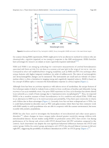
Figure 2. Absorption coefficient of Oxy-hemoglobin (HbO), Deoxy-hemoglobin (HbR) and water in the near infrared region
the scanner during fMRI experiments, fNIRS might prove to be an alternative method for subjects who are
claustrophobic, cognitive impaired, or too young to cooperate in the MRI environment. fNIRS therefore
have advantages for research on infants or more cognitively impaired individuals [92-95] .
NIRS and fNIRS is an emerging technology for noninvasive measurements of cerebral hemodynamics
associated with brain activity. It is not done in a scanner, and uses light in the range of 700 nm to 1000 nm.
Compared to other well-established brain imaging modalities, such as fMRI and PET, this technique offers
unique features with higher temporal resolution (in order of milliseconds). The status of oxyhemoglobin
and deoxyhemoglobin changes can be measured. The instruments are small and are tolerant of subject
motion which is often a limitation in imaging young and cognitively impaired subjects. fNIRS is based on
the concept of diffuse optics to measure the hemodynamic response in cortical regions [Figure 2].
Although there has been no previous experience with inborn errors of metabolism, the unique features of
this technique makes it ideal to evaluate brain activity in these conditions at baseline and ultimately during
recovery of an acute metabolic event. Our prior fMRI experience in Urea cycle disorders has shown altered
[91]
brain networks as a result of brain damage due to hyperammonemic encephalopathy . Thus, we expected
fNIRS to be a sensitive measure of brain hemodynamics in this condition and related IEMs. fNIRS may
be an alternative to fMRI due to its noninvasiveness and portability. It has been used extensively in infants
and children due to these advantages [Figure 3]. Currently there has not been widespread use in IEMs, but
it could find potential in disorders such as PKU and galactosemia where there has been extensive work
with fMRI mainly in adult populations who are able to cooperate with the scanner environment, but more
recently with pediatric populations.
fNIRS has also been used to investigate the biomarkers of mitochondrial and other neuro-genetic
[95]
disorders , where changes in tissue oxygen index showed greater variability among children with
mitochondrial disease. Recent studies using fNIRS on prefrontal cortex (PFC) have shown that during
performance of the Stroop task cohorts with OTCD showed lower left PFC activation compared to the
controls. Such observation was despite the non-significant behavioral differences between the OTCD and
control group. Unlike the control group, participants with OTCD also showed bilateral increase in left and
[96]
right PFC, suggesting the possibility of inefficient lateralization of PFC in OTCD group [Figure 4] .